


 膜杰作
膜杰作 Star Staining


分子别名(Synonym)
PDCD1,PD1,CD279,SLEB2
表达区间及表达系统(Source)
Human PD-1, His Tag (PD1-H5221) is expressed from human 293 cells (HEK293). It contains AA Leu 25 - Gln 167 (Accession # NP_005009.2).
Predicted N-terminus: Leu 25
Request for sequence
蛋白结构(Molecular Characterization)

This protein carries a polyhistidine tag at the C-terminus.
The protein has a calculated MW of 16.8 kDa. The protein migrates as 33-38 kDa when calibrated against Star Ribbon Pre-stained Protein Marker under reducing (R) condition (SDS-PAGE) due to glycosylation.
内毒素(Endotoxin)
Less than 1.0 EU per μg by the LAL method.
纯度(Purity)
>95% as determined by SDS-PAGE.
制剂(Formulation)
Lyophilized from 0.22 μm filtered solution in PBS, pH7.4 with trehalose as protectant.
Contact us for customized product form or formulation.
重构方法(Reconstitution)
Please see Certificate of Analysis for specific instructions.
For best performance, we strongly recommend you to follow the reconstitution protocol provided in the CoA.
存储(Storage)
For long term storage, the product should be stored at lyophilized state at -20°C or lower.
Please avoid repeated freeze-thaw cycles.
This product is stable after storage at:
- -20°C to -70°C for 24 months in lyophilized state;
- -70°C for 12 months under sterile conditions after reconstitution.
质量管理控制体系(QMS)
电泳(SDS-PAGE)
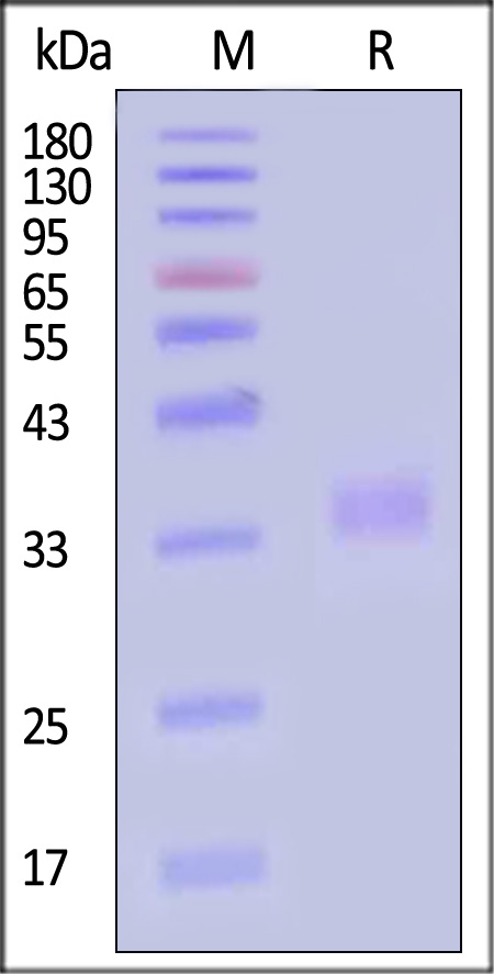
Human PD-1, His Tag on SDS-PAGE under reducing (R) condition. The gel was stained with Coomassie Blue. The purity of the protein is greater than 95% (With Star Ribbon Pre-stained Protein Marker).
SEC-MALS
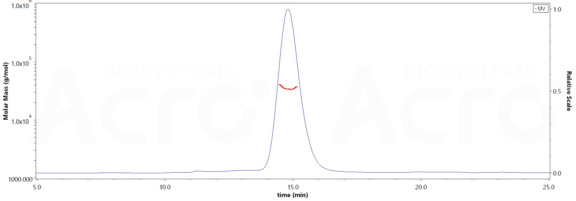
The purity of Human PD-1, His Tag (Cat. No. PD1-H5221) is more than 85% and the molecular weight of this protein is around 28-42 kDa verified by SEC-MALS.
Report
活性(Bioactivity)-ELISA
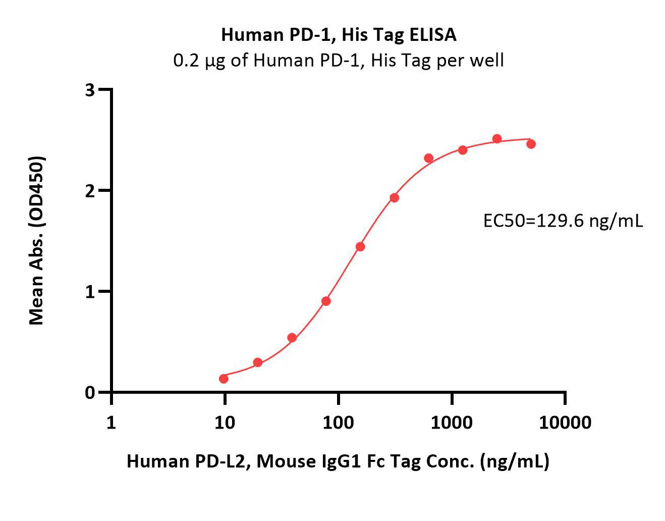
Immobilized Human PD-1, His Tag (Cat. No. PD1-H5221) at 2 μg/mL (100 μL/well) can bind Human PD-L2, Mouse IgG1 Fc Tag (Cat. No. PD2-H52A5) with a linear range of 10-156 ng/mL (Routinely tested).
Protocol
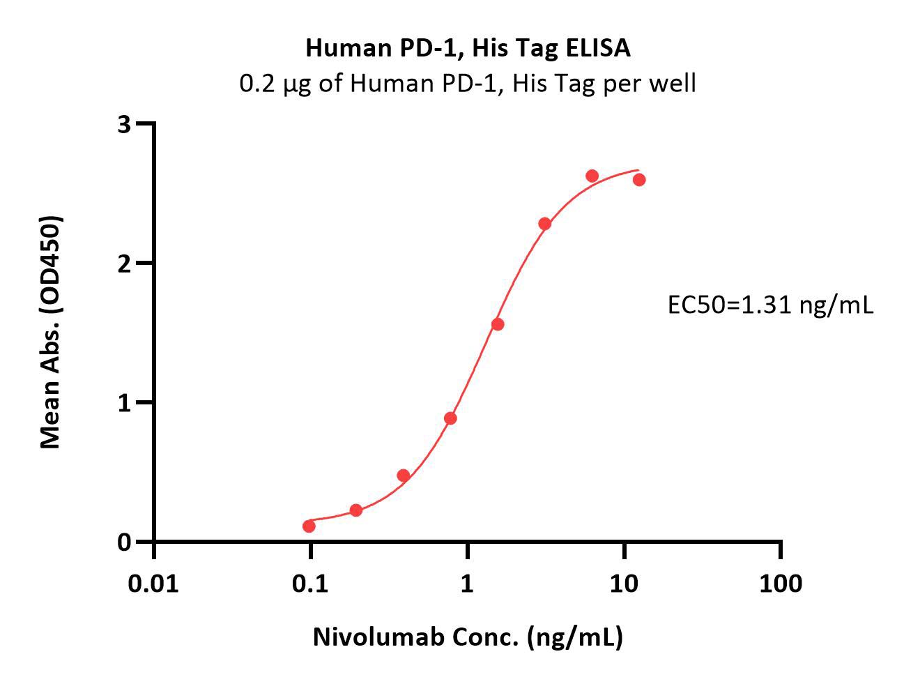
Immobilized Human PD-1, His Tag (Cat. No. PD1-H5221) at 2 μg/mL (100 μL/well) can bind Nivolumab with a linear range of 0.1-3 ng/mL (Routinely tested).
Protocol
活性(Bioactivity)-SPR
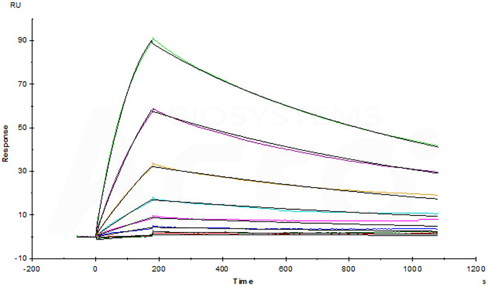
Opdivo (Nivolumab) captured on CM5 chip via anti-human IgG Fc antibodies surface, can bind Human PD-1, His Tag (Cat. No. PD1-H5221) with an affinity constant of 4.94 nM as determined in a SPR assay (Biacore T200) (Routinely tested).
Protocol
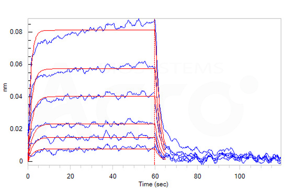
活性(Bioactivity)-BLI
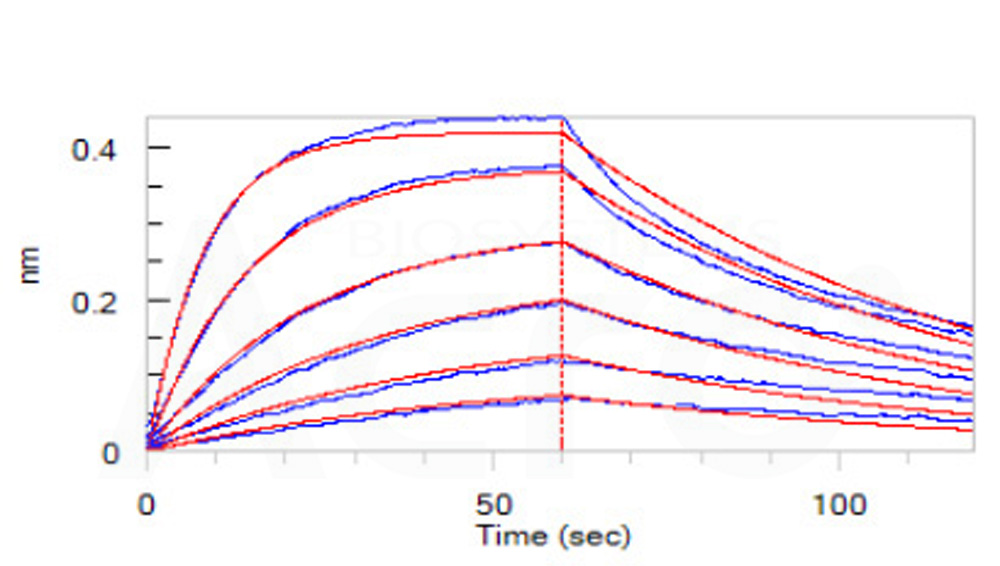
Loaded Human PD-1, His Tag (Cat. No. PD1-H5221) on HIS1K Biosensor, can bind Human PD-L1, Fc Tag (HPLC verified) (Cat. No. PD1-H5258) with an affinity constant of 38.9 nM as determined in BLI assay (ForteBio Octet Red96e) (QC tested).
Protocol

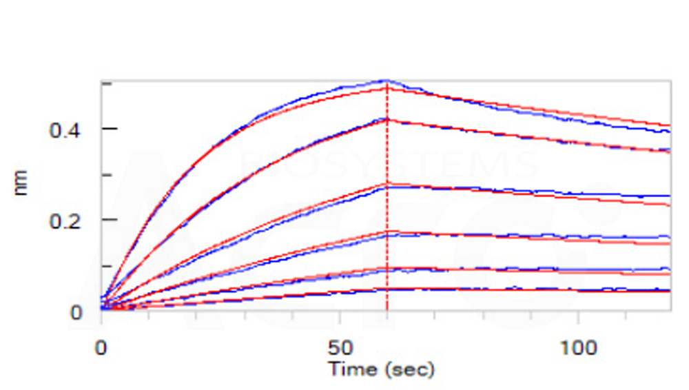
Loaded Human PD-1, His Tag (Cat. No. PD1-H5221) on HIS1K Biosensor, can bind Human PD-L2, Fc Tag (HPLC verified) (Cat. No. PD2-H5251) with an affinity constant of 16.3 nM as determined in BLI assay (ForteBio Octet Red96e) (QC tested).
Protocol
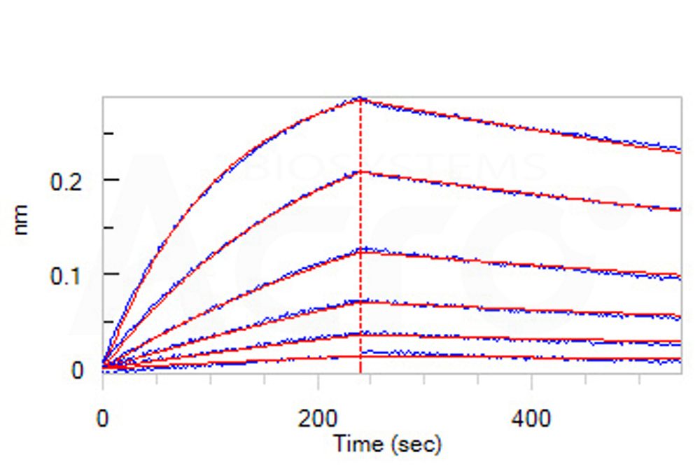
Loaded Opdivo (Nivolumab) on ProteinA Biosensor, can bind Human PD-1, His Tag (Cat. No. PD1-H5221) with an affinity constant of 5.09 nM as determined in BLI assay (ForteBio Octet Red96e) (Routinely tested).
Protocol
Loaded Human PD-L1, Mouse IgG1 Fc Tag, low endotoxin (HPLC-verified) (Cat. No. PD1-H52A3) on AMC Biosensor, can bind Human Human PD-1, His Tag (Cat. No. PD1-H5221) with an affinity constant of 2.1 μM as determined in BLI assay (ForteBio Octet Red96e) (Routinely tested).
Protocol
 +添加评论
+添加评论


产品推荐(Recommended Products)

背景(Background)
Programmed cell death protein 1 (PD-1) is also known as CD279 and PDCD1, is a type I membrane protein and is a member of the extended CD28/CTLA-4 family of T cell regulators. PDCD1 is expressed on the surface of activated T cells, B cells, macrophages, myeloid cells and a subset of thymocytes. PD-1 has two ligands, PD-L1 and PD-L2, which are members of the B7 family. PD-L1 is expressed on almost all murine tumor cell lines, including PA1 myeloma, P815 mastocytoma, and B16 melanoma upon treatment with IFN-γ. PD-L2 expression is more restricted and is expressed mainly by DCs and a few tumor lines. PD1 inhibits the T-cell proliferation and production of related cytokines including IL-1, IL-4, IL-10 and IFN-γ by suppressing the activation and transduction of PI3K/AKT pathway. In addition, coligation of PD1 inhibits BCR-mediating signal by dephosphorylating key signal transducer. In vitro, treatment of anti-CD3 stimulated T cells with PD-L1-Ig results in reduced T cell proliferation and IFN-γ secretion. Monoclonal antibodies targeting PD-1 that boost the immune system are being developed for the treatment of cancer.


