登录 | 注册    关注公众号  
微信公众号
搜索
 >  Protein>FGF R4 >FG4-H5228

Human FGF R4 / CD334 Protein, His Tag

热销产品推荐:

ACROBiosystems提供针对多种生物标志物靶标的lHC抗体产品,具有:验证充分、质量可靠、灵活授权及免疫组化检测服务支持!

分子别名(Synonym)

FGFR4,CD334,JTK2,MGC20292,TKF

表达区间及表达系统(Source)

Human FGF R4, His Tag (FG4-H5228) is expressed from human 293 cells (HEK293). It contains AA Leu 22 - Asp 369 (Accession # P22455-1).

Predicted N-terminus: Leu 22

Request for sequence

蛋白结构(Molecular Characterization)

FGF R4 Structure

This protein carries a polyhistidine tag at the C-terminus.

The protein has a calculated MW of 39.3 kDa. The protein migrates as 50-70 kDa under reducing (R) condition (SDS-PAGE) due to glycosylation.

内毒素(Endotoxin)

Less than 1.0 EU per μg by the LAL method.

纯度(Purity)

>95% as determined by SDS-PAGE.

制剂(Formulation)

Lyophilized from 0.22 μm filtered solution in PBS, pH7.4 with trehalose as protectant.

Contact us for customized product form or formulation.

重构方法(Reconstitution)

Please see Certificate of Analysis for specific instructions.

For best performance, we strongly recommend you to follow the reconstitution protocol provided in the CoA.

存储(Storage)

For long term storage, the product should be stored at lyophilized state at -20°C or lower.

Please avoid repeated freeze-thaw cycles.

This product is stable after storage at:

  1. -20°C to -70°C for 12 months in lyophilized state;
  2. -70°C for 3 months under sterile conditions after reconstitution.

质量管理控制体系(QMS)

  1. 质量管理体系(ISO, GMP)
  2. 质量优势
  3. 质控流程
 

电泳(SDS-PAGE)

FGF R4 SDS-PAGE

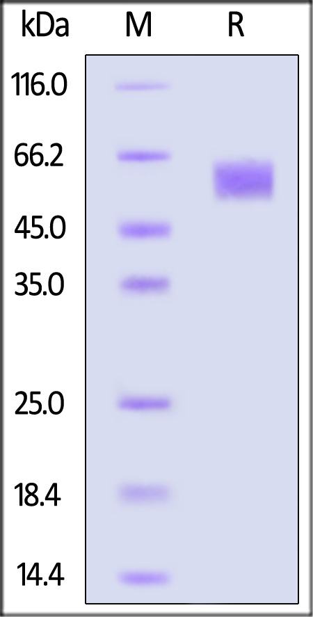
Human FGF R4, His Tag on SDS-PAGE under reducing (R) condition. The gel was stained with Coomassie Blue. The purity of the protein is greater than 95%.

 
评论(0)
  1. 182XXXXXXX4
  2. 6人赞
  3. This protein has a particularly good affinity and is relatively stable. It is dissolved under recommended conditions, and there is little difference between batches when used. In ELISA experiments, it has good reproducibility whether used as a coating or as a standard substance. protocal is very detailed, there are giveaways, logistics is fast
  4. 2023-4-12
  1. 651XXXXXXX
  2. 3人赞
  3. The c-MET protein was delivered on time and we were able to use this protein for ELISA screening. I was able to get really good results from using this protein and would come back to Acrobiosystems to reorder this protein or order other proteins that I will need for future experiments.
  4. 2022-6-10
  1. 188XXXXXXX9
  2. 1人赞
  3. 此蛋白亲和力特别好,而且比较稳定,在推荐的条件下进行溶解处理,使用时不同批次之间差异很小,在ELISA实验中,不管是用于包被还是标准品都有很好的重现性,后续还会继续使用,而且也会尝试其他蛋白。protocal也很详细,还有赠品,物流也快
  4. 2023-3-19
 
ACRO质量管理体系


背景(Background)

Fibroblast growth factor receptor 4(FGFR4) is also known as CD334, JTK2, hydroxyaryl-protein kinase, TKF, protein-tyrosine kinase . The FGFR4 gene provides instructions for making a protein called fibroblast growth factor receptor 4. This protein is part of a family of fibroblast growth factor receptors that share similar structures and functions. These receptor proteins play a role in important processes such as cell division, regulating cell growth and maturation, formation of blood vessels, wound healing, and embryo development.The FGFR4 protein interacts with specific growth factors to conduct signals from the environment outside the cell to the nucleus. The nucleus responds to these signals by switching on or off appropriate genes that help the cell adjust to changes in the environment. In response, the cell might divide, move, or mature to take on specialized functions. Although specific functions of FGFR4 remain unclear, studies indicate that the gene is involved in muscle development and the maturation of bone cells in the skull. The FGFR4 gene may also play a role in the development and maintenance of specialized cells (called foveal cones) in the light-sensitive layer (the retina) at the back of the eye.


前沿进展

Optimal injection sites for therapeutic angiogenesis: HGF-mediated regulation of HIF-1α via MAPK/PI3K pathways in hypoxic endothelial cells
Wang, Rong, Li et al
Tissue Cell (2025) 95, 102871
Abstract: Therapeutic angiogenesis offers a promising strategy for patients with critical limb-threatening ischemia (CLTI) who are unsuitable candidates for revascularization. However, the optimal administration sites for gene therapy agents, such as pCK-HGF-X7, remains undefined. Clinical trials commonly employ multiple intramuscular injections at sites of arterial occlusion; yet the necessity and efficacy of such extensive and repetitive protocols remains unclear. Targeted injections into ischemic tissues or their margins may improve therapeutic outcomes. Moreover, the molecular mechanisms by which hepatocyte growth factor (HGF)/c-Met signaling regulates hypoxia-inducible factor-1α (HIF-1α) expression under hypoxic conditions are not fully understood. This study aims to elucidate these molecular mechanisms in endothelial cells under hypoxic conditions and to identify the most effective injection sites for therapeutic angiogenesis agents. The effects of various HGF isoforms/complexes on human aortic endothelial cells (HAECs) were evaluated under normoxic and hypoxic conditions, focusing on proliferation, migration, and tube formation. Pathway inhibitors were used to explore the underlying mechanisms in hypoxic HAECs, and the findings were validated in a rat hindlimb ischemia model. Results demonstrated that HGF723 and HGF728 activated the mitogen-activated protein kinase (MAPK) and phosphatidylinositol 3-kinase (PI3K) pathways, regulating HIF expression and significantly enhanced endothelial cell proliferation, migration, and tube formation, particularly under hypoxia. Despite these cellular effects, HGF treatment did not significantly improve tissue perfusion or neovascularization in normal rat hindlimbs. However, in ischemic rat hindlimbs, it markedly promoted angiogenesis and improved tissue perfusion in the gastrocnemius muscle. These findings indicate that therapeutic angiogenesis agents should primarily target hypoxic tissues, extending to the interface between normoxic and hypoxic regions, to optimize treatment efficacy.Copyright © 2025 Elsevier Ltd. All rights reserved.
Effects of Chlorella protothecoides-derived polydeoxyribonucleotides on skin regeneration and wound healing
Park, Nam, Lee et al
Arch Dermatol Res (2025) 317 (1), 483
Abstract: The skin acts as a crucial barrier and, upon injury, initiates complex wound-healing processes involving various cell types. Polydeoxyribonucleotides (PDRNs) are well-known for their efficacy in enhancing skin regeneration and wound healing. This study sought to investigate the effectiveness of PDRNs derived from Chlorella protothecoides, a sustainable and scalable microalgal source, in promoting skin regeneration and wound healing. Keratinocytes and fibroblasts were used for assessing the impact of PDRNs on cell proliferation, migration, collagen synthesis, and angiogenesis. Gene expression and associated signaling pathways were also examined using RT-qPCR and Western blot analyses. Our findings demonstrated that PDRNs significantly enhanced the proliferation and migration of skin cells, upregulated growth arrest specific 6 (GAS6) and hepatocyte growth factor (HGF) expression, and increased collagen synthesis by modulating collagen type I alpha 1 (COLIA1) expression. Additionally, PDRNs enhanced angiogenesis by promoting vascular endothelial growth factor (VEGF) expression and activation of ERK, AKT, β-catenin and STAT3 pathways via an adenosine A2A receptor (A2AR)-dependent mechanism. These findings suggest that microalgal-derived PDRNs have significant potential as sustainable and effective agents for clinical and cosmetic applications aimed at improving skin health and wound healing.© 2025. The Author(s), under exclusive licence to Springer-Verlag GmbH Germany, part of Springer Nature.
Genetic insights into avian influenza resistance in Jeju Island chickens: the roles of Mx1 and oligoadenylate synthetase-like single nucleotide polymorphisms
Kim, Jeong, Yang et al
J Anim Sci Technol (2025) 67 (1), 69-85
Abstract: Influenza A virus (FLUAV) causes serious diseases in both poultry and humans. Various host proteins, including Mx1, are considered candidates for avian influenza (AI) resistance. After infecting Jeju Native chicken embryo fibroblasts (CEFs) with three types of AI viruses, we performed gene expression profiling, identified single nucleotide polymorphisms (SNPs) through RNA-sequencing, and confirmed phenotypes showing antiviral activity in vitro. Highly pathogenic AI viruses upregulated FGF2, LYN, and FLT4 and downregulated HGF, ANGPT1, and ROR2, while a low pathogenicity AI upregulated PARK7, RACK1, and DTX3L and downregulated SIRT1, LRRK2, and WAC. However, no virus affected Mx1 expression. Although SNPs in Mx1 could not discriminate antiviral activity alone, the only CEF resistant to H5N6, strain AN4, contained the Mx1 631 R/R genotype and strongly expressed an oligoadenylate synthetase-like (OASL) variant with a unique SNP: c.G880A (p.E294K). Using transfected cell lines, H5N6-infected cells expressing OASL with the c.G880A SNP showed minimal cytopathic effects and the lowest M gene expression. This study confirms that Jeju Native chickens with specific SNP combinations in both Mx1 and OASL showed H5N6 resistance and demonstrates the interplay of genetic factors in host-pathogen dynamics, suggesting a need for integrated analyses of multiple resistance genes to inform AI prevention strategies.© Copyright 2025 Korean Society of Animal Science and Technology.
Super-Resolution Fluorescence Imaging Reveals the Mechanism of NRP1 Clustering on Non-Small-Cell Lung Cancer Membranes
Li, Gao, Qi et al
Anal Chem (2025) 97 (4), 2326-2334
Abstract: Neuropilin 1 (NRP1) is upregulated in various types of malignant tumors, especially non-small-cell lung cancer (NSCLC). However, the precise mechanisms for membrane localization and regulation are not fully understood. Observations from super-resolution microscopy have revealed that NRP1 tends to form nanoscale clusters on the cell membrane, with these clusters varying significantly in size and density across different regions. Further research has shown that stimulation by hepatocyte growth factor (HGF) can reorganize the distribution of NRP1, reducing the number of small clusters while promoting the formation of larger ones. This suggests a propensity for internalization after activation. Additionally, dual-color dSTORM imaging has demonstrated a certain degree of colocalization between NRP1 and c-MET, indicating that c-MET plays an important role in stabilizing NRP1 clusters. This study provides new insights into the mechanism behind NRP1's clustered distribution on cell membranes and paves the way for developing more effective therapeutic strategies targeting NRP1 within tumors.
Showing 1-4 of 810 papers.
Powered by BizGenius
 
 
货号/价格
文档
联系电话:
+86 400-682-2521(全国)
010-53681107(北京)
021-50850665(上海)
运输方式
订单邮箱:
order.cn@acrobiosystems.com
技术支持邮箱:
tech.cn@acrobiosystems.com
FGF R4靶点信息
英文全称:Fibroblast growth factor receptor 4
中文全称:成纤维细胞生长因子受体-4
种类:Homo sapiens
上市药物数量:3详情
临床药物数量:17详情
最高研发阶段:批准上市
查看更多信息
前沿进展
点击查看详细

消息提示

请输入您的联系方式,再点击提交!

确定