登录 | 注册    关注公众号  
微信公众号
搜索
 >  Protein>Oncostatin M >OSM-H5213

Human Oncostatin M (OSM) Protein, premium grade

分子别名(Synonym)

OSM,MGC20461,Oncostatin M

表达区间及表达系统(Source)

Human Oncostatin M Protein, premium grade (OSM-H5213) is expressed from human 293 cells (HEK293). It contains AA Ala 26 - Arg 252 (Accession # NP_065391.1).

Predicted N-terminus: Ala 26

It is produced under our rigorous quality control system that incorporates a comprehensive set of tests including sterility and endotoxin tests. Product performance is carefully validated and tested for compatibility for cell culture use or any other applications in the early preclinical stage. When ready to transition into later clinical phases, we also offer a custom GMP protein service that tailors to your needs. We will work with you to customize and develop a GMP-grade product in accordance with your requests that also meets the requirements for raw and ancillary materials use in cell manufacturing of cell-based therapies.

Request for sequence

蛋白结构(Molecular Characterization)

Oncostatin M Structure

This protein carries no "tag".

The protein has a calculated MW of 25.8 kDa. The protein migrates as 36 kDa under reducing (R) condition (SDS-PAGE) due to glycosylation.

内毒素(Endotoxin)

Less than 0.1 EU per μg by the LAL method.

无菌(Sterility)

Negative

纯度(Purity)

>95% as determined by SDS-PAGE.

制剂(Formulation)

Lyophilized from 0.22 μm filtered solution in PBS, pH7.4 with trehalose as protectant.

Contact us for customized product form or formulation.

重构方法(Reconstitution)

Please see Certificate of Analysis for specific instructions.

For best performance, we strongly recommend you to follow the reconstitution protocol provided in the CoA.

存储(Storage)

For long term storage, the product should be stored at lyophilized state at -20°C or lower.

Please avoid repeated freeze-thaw cycles.

This product is stable after storage at:

  1. -20°C to -70°C for 12 months in lyophilized state;
  2. -70°C for 3 months under sterile conditions after reconstitution.

质量管理控制体系(QMS)

  1. 质量管理体系(ISO, GMP)
  2. 质量优势
  3. 质控流程
 

电泳(SDS-PAGE)

Oncostatin M SDS-PAGE

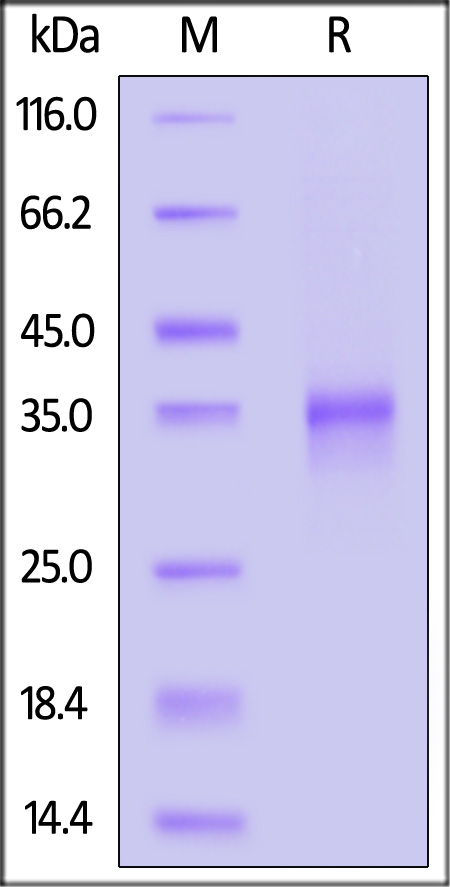
Human Oncostatin M Protein, premium grade on SDS-PAGE under reducing (R) condition. The gel was stained with Coomassie Blue. The purity of the protein is greater than 95%.


活性(Bioactivity)-ELISA

Oncostatin M ELISA

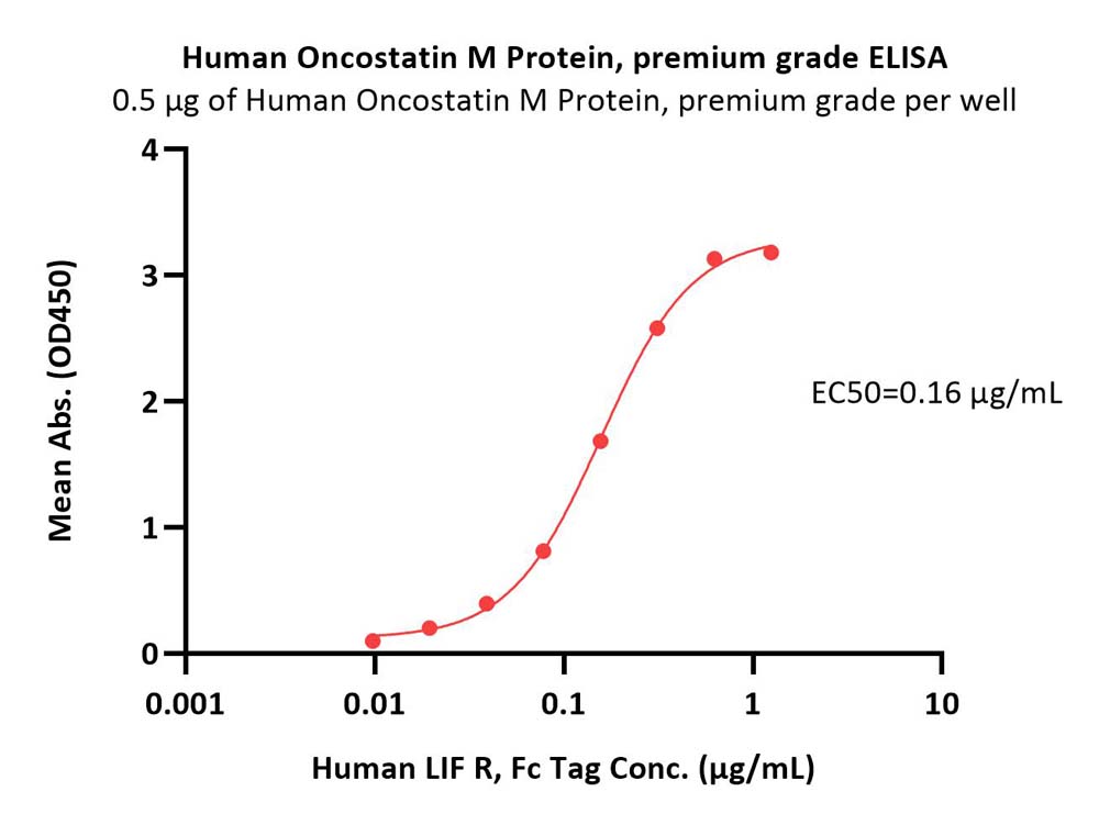
Immobilized Human Oncostatin M Protein, premium grade (Cat. No. OSM-H5213) at 5 μg/mL (100 μL/well) can bind Human LIF R, Fc Tag (Cat. No. LIR-H4252) with a linear range of 0.01-0.312 μg/mL (QC tested).

Protocol

 
评论(0)
 
ACRO质量管理体系


背景(Background)

Oncostatin M is also known as OSM, is a glycoprotein belonging to the interleukin-6 family of cytokines that has functions mainly in cell growth. Of these cytokines it most closely resembles leukemia inhibitory factor (LIF) in both structure and function. However, it is as yet poorly defined and is proving important in liver development, haematopoeisis, inflammation and possibly CNS development. It is also associated with bone formation and destruction. OSM signals through cell surface receptors that contain the protein gp130. The type I receptor is composed of gp130 and LIFR, the type II receptor is composed of gp130 and OSMR. Oncostatin M (OSM) was previoustly identified by its ability to inhibit the growth of cells from melanoma and other solid tumors. It also has been reported that OSM, like LIF, IL-6 and G-CSF, has the ability to inhibit the proliferation of murine M1 myeloid leukemic cells and can induce their differentiation into macrophage-like cells. The human form of OSM is insensitive between pH2 and 11 and resistant to heating for one hour at 56 degree but is not stable at 90 degrees. The three dimensional structure of human OSM has been solved to atomic resolution, confirming the predicted long chain four helix bundle topology. Comparing this structure with the known structures of other known LC cytokines shows it to be most closely related to LIF.


前沿进展

Sensitivity to Neutralizing Antibodies and Resistance to Type I Interferons in SARS-CoV-2 R.1 Lineage Variants, Canada
Jacob, Zhang, Ajoge et al
Emerg Infect Dis (2023) 29 (7), 1386-1396
Abstract: Isolating and characterizing emerging SARS-CoV-2 variants is key to understanding virus pathogenesis. In this study, we isolated samples of the SARS-CoV-2 R.1 lineage, categorized as a variant under monitoring by the World Health Organization, and evaluated their sensitivity to neutralizing antibodies and type I interferons. We used convalescent serum samples from persons in Canada infected either with ancestral virus (wave 1) or the B.1.1.7 (Alpha) variant of concern (wave 3) for testing neutralization sensitivity. The R.1 isolates were potently neutralized by both the wave 1 and wave 3 convalescent serum samples, unlike the B.1.351 (Beta) variant of concern. Of note, the R.1 variant was significantly more resistant to type I interferons (IFN-α/β) than was the ancestral isolate. Our study demonstrates that the R.1 variant retained sensitivity to neutralizing antibodies but evolved resistance to type I interferons. This critical driving force will influence the trajectory of the pandemic.
Downregulation of SOCS gene expression can inhibit the formation of acute and persistent BDV infections
Li, Xia, Meng et al
Scand J Immunol (2021) 93 (1), e12974
Abstract: High expression of suppressors of cytokine signalling (SOCS) has been detected during various viral infections. As a negative feedback regulator, SOCS participates in the regulation of multiple signalling pathways. In this study, to study the related mechanism between SOCS and BDV and to explore the effect of SOCS on IFN pathways in nerve cells, downregulated of SOCS1/3 in oligodendroglial (OL) cells and OL cells persistently infected with BDV (OL/BDV) were constructed with RNA interference technology. An interferon inducer (poly I:C, PIC) and an IFN-α/β R1 antibody were used as stimulation in the SOCS1/3 low-expression cell models, qRT-PCR was used to detect type I IFN and BDV nucleic acid expression, Western blot was used to detect the expression of BDV P40 protein. After BDV acute infection with OL cells which with downregulated SOCS expression, the virus accounting was not detected, and the viral protein expression was lower than that of OL/BDV cells; the OL/BDV cells with downregulated SOCS expression had lower virus nucleic acid and protein expression than OL/BDV cells. Stimulated by IFN-α/β R1 antibody, the expression of type I interferon in OL/BDV cells decreased, and the content of BDV nucleic acid and protein increased, which was higher than that of OL/BDV cells. From the results, it was concluded that downregulating SOCS1/3 can inhibit the formation of acute BDV infection and virus replication in persistent BDV infection by promoting the expression of IFN-α/β and that SOCS can be used as a new target for antiviral therapy.© 2020 The Scandinavian Foundation for Immunology.
Glucocorticoid regulation of 24-hour oscillation in interferon receptor gene expression in mouse liver
Koyanagi, Suyama, Kuramoto et al
Endocrinology (2006) 147 (11), 5034-40
Abstract: Although the antiviral effect of interferon (IFN) varies depending on 24-h oscillation in the expression of its specific receptor, the mechanism of oscillation remains to be clarified. Here we report that oscillation in the expression of the IFN receptor gene (IFN-alpha/beta R1) in mouse liver is caused by the endogenous rhythm of glucocorticoid secretion. Brief exposure of mouse hepatic cells (Hepa 1-6) to corticosterone (CORT) resulted in a significant decrease in mRNA levels of IFN-alpha/beta R1. The CORT-induced decrease in IFN-alpha/beta R1 mRNA levels was reversed by pretreating the cells with RU486, a glucocorticoid receptor antagonist. The mRNA levels of IFN-alpha/beta R1 gene in the liver of adrenalectomized mice were consistently increased throughout the day. However, a single administration of CORT to adrenalectomized mice significantly decreased the mRNA levels of IFN-alpha/beta R1 in the liver. Furthermore, the rhythmic phase of IFN-alpha/beta R1 expression was modulated after the alteration of rhythmicity in glucocorticoid secretion, which was induced by restricted daily feeding. As a consequence, under manipulation of the feeding schedule, 2'-5' oligoadenylate synthase activities, as an index of antiviral effect, in plasma and liver at 24 h after IFN-alpha injection also varied depending on the alteration of glucocorticoid secretion rhythm. These results suggest that the endogenous rhythm of glucocorticoid secretion is involved in the circadian regulation of IFN-alpha/beta R1 expression in mouse liver. Our findings also support the notion that monitoring the 24-h variation in IFN receptor function is useful for selecting the most appropriate time of day to administer IFN.
Role of interferon alpha/beta receptor chain 1 in the structure and transmembrane signaling of the interferon alpha/beta receptor complex
Constantinescu, Croze, Wang et al
Proc Natl Acad Sci U S A (1994) 91 (20), 9602-6
Abstract: A previously cloned cDNA encodes one subunit of the human interferon alpha/beta receptor (IFN alpha R), denoted IFN alpha R1. To study the expression and signaling of IFN alpha R1, we used monoclonal antibodies (mAbs) generated against the baculovirus-expressed ectodomain of IFN alpha R1. Immunoprecipitation and immunoblotting of lysates from a variety of human cell lines showed that IFN alpha R1 has an apparent molecular mass of 135 kDa. Binding analysis with 125I-labeled mAb demonstrated high levels of cell surface expression of IFN alpha R1 in human cells and in mouse cells transfected with IFN alpha R1 cDNA, whereas no cross-reactivity was observed in control mouse L929 cells expressing only the endogenous mouse receptor. The subunit was rapidly down-regulated by IFN alpha (80% decrease within 2 hr) and degraded upon internalization. The IFN alpha R1 chain appeared to be constitutively associated with the 115-kDa subunit of the IFN alpha/beta receptor, since the mAbs coprecipitated this protein. IFN alpha/beta treatment induced tyrosine phosphorylation of IFN alpha R1 within 1 min, with kinetics paralleling that of the IFN-activated protein-tyrosine kinases Jak1 and Tyk2. Ligand-induced tyrosine phosphorylation of IFN alpha R1 was blocked by the kinase inhibitors genistein or staurosporine. Although IFN alpha R1 cDNA-transfected mouse cells expressed high levels of this subunit when compared with empty vector-transfected cells the number of binding sites for human IFN alpha (50-75 sites per cell) was not increased. Human IFN alpha induced the expression of a mouse IFN alpha/beta-responsive gene (the 204 gene) in mouse L929 cells transfected with the IFN alpha R1 cDNA, but not in mock-transfected cells. These results suggest that the IFN alpha R1 subunit acts as a species-specific signal transduction component of the IFN alpha/beta receptor complex.
Showing 1-4 of 5 papers.
Powered by BizGenius
 
 
货号/价格
文档
联系电话:
+86 400-682-2521(全国)
010-53681107(北京)
021-50850665(上海)
运输方式
订单邮箱:
order.cn@acrobiosystems.com
技术支持邮箱:
tech.cn@acrobiosystems.com
Oncostatin M靶点信息
英文全称:Oncostatin M
中文全称:肿瘤抑制因子
种类:Homo sapiens
上市药物数量:0详情
临床药物数量:1详情
最高研发阶段:临床二期
查看更多信息
前沿进展
点击查看详细

消息提示

请输入您的联系方式,再点击提交!

确定