登录 | 注册    关注公众号  
微信公众号
搜索
 >  Protein>FOLR1 >FO1-M82E9

Biotinylated Mouse FOLR1 Protein, His,Avitag™ (MALS verified)

分子别名(Synonym)

FOLR-1,FBP,FOLR,FRα

表达区间及表达系统(Source)

Biotinylated Mouse FOLR1, His,Avitag (FO1-M82E9) is expressed from human 293 cells (HEK293). It contains AA Thr 25 - Ser 232 (Accession # P35846-1).

Predicted N-terminus: Thr 25

Request for sequence

蛋白结构(Molecular Characterization)

FOLR1 Structure

This protein carries a polyhistidine tag at the C-terminus, followed by an Avi tag (Avitag™).

The protein has a calculated MW of 26.9 kDa. The protein migrates as 32-45 kDa under reducing (R) condition (SDS-PAGE) due to glycosylation.

标记(Labeling)

Biotinylation of this product is performed using Avitag™ technology. Briefly, the single lysine residue in the Avitag is enzymatically labeled with biotin.

蛋白标记度(Protein Ratio)

Passed as determined by the HABA assay / binding ELISA.

内毒素(Endotoxin)

Less than 1.0 EU per μg by the LAL method.

纯度(Purity)

>95% as determined by SDS-PAGE.

>90% as determined by SEC-MALS.

制剂(Formulation)

Lyophilized from 0.22 μm filtered solution in PBS, pH7.4 with trehalose as protectant.

Contact us for customized product form or formulation.

重构方法(Reconstitution)

Please see Certificate of Analysis for specific instructions.

For best performance, we strongly recommend you to follow the reconstitution protocol provided in the CoA.

存储(Storage)

For long term storage, the product should be stored at lyophilized state at -20°C or lower.

Please avoid repeated freeze-thaw cycles.

This product is stable after storage at:

  1. -20°C to -70°C for 12 months in lyophilized state;
  2. -70°C for 3 months under sterile conditions after reconstitution.

质量管理控制体系(QMS)

  1. 质量管理体系(ISO, GMP)
  2. 质量优势
  3. 质控流程
 

电泳(SDS-PAGE)

FOLR1 SDS-PAGE

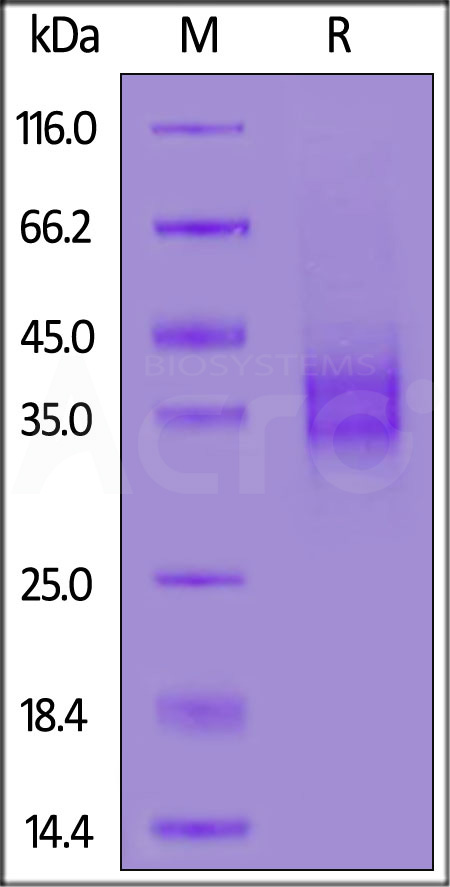
Biotinylated Mouse FOLR1, His,Avitag on SDS-PAGE under reducing (R) condition. The gel was stained with Coomassie Blue. The purity of the protein is greater than 95%.

SEC-MALS

FOLR1 SEC-MALS

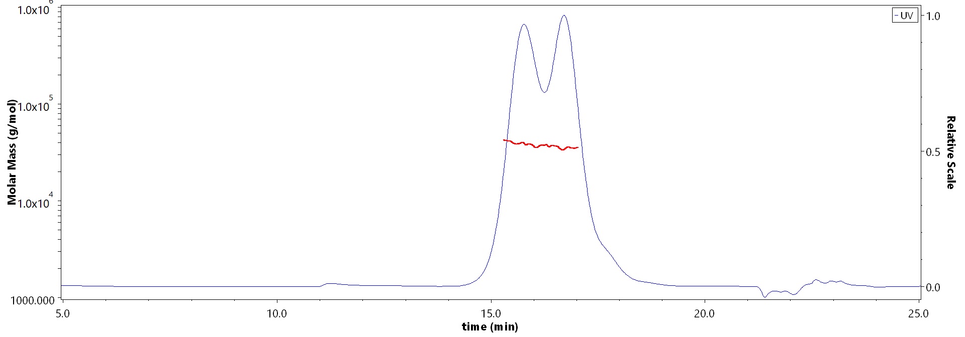
The purity of Biotinylated Mouse FOLR1, His,Avitag (Cat. No. FO1-M82E9) is more than 90% and the molecular weight of this protein is around 30-45 kDa verified by SEC-MALS.

Report


活性(Bioactivity)-ELISA

FOLR1 ELISA

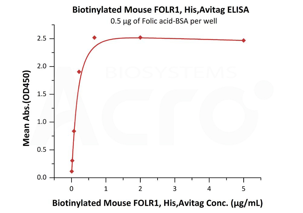
Immobilized Folic acid-BSA at 5 μg/mL (100 μL/well) can bind Biotinylated Mouse FOLR1, His,Avitag (Cat. No. FO1-M82E9) with a linear range of 0.008-0.222 μg/mL (QC tested).

Protocol

 
评论(0)
 
ACRO质量管理体系


背景(Background)

Folate Receptor 1 (FOLR1) is also known as Folate receptor alpha, Folate Binding Protein (FBP), FOLR, and is a member of the folate receptor (FOLR) family. Members of this gene family have a high affinity for folic acid and for several reduced folic acid derivatives, and mediate delivery of 5-methyltetrahydrofolate to the interior of cells. Mature FOLR1 is an N-glycosylated protein that is anchored to the cell surface by a GPI linkage. FOLR1 is predominantly expressed on epithelial cells and is dramatically upregulated on many carcinomas. FOLR1 is internalized to the endosomal system where it dissociates from its ligand before recycling to the cell surface. A soluble form of FOLR1 can be proteolytically shed from the cell surface into the serum and breast milk. Defects in FOLR1 are the cause of neurodegeneration due to cerebral folate transport deficiency (NCFTD). NCFTD is an autosomal recessive disorder resulting from brain-specific folate deficiency early in life.


前沿进展

Patient outcomes in advanced ovarian cancer treated with an anti-FOLR1 antibody-drug conjugate
Johannet, Flint, Chui et al
Gynecol Oncol (2025) 195, 173-179
Abstract: Mirvetuximab soravtansine-gynx (MIRV) is a FOLR1-binding antibody-drug conjugate (ADC) with a microtubule inhibitor payload. We investigated MIRV's efficacy, toxicity profile, and determinants of resistance in a cohort of patients with recurrent/persistent high FOLR1-expressing high-grade serous ovarian cancer (HGSOC).This retrospective study included 170 patients with recurrent/persistent FOLR1-high (≥75 % of tumor cells with ≥2+ membranous staining intensity) HGSOC who received standard-of-care MIRV monotherapy. We evaluated progression-free survival (PFS) and overall survival (OS) using the Kaplan-Meier method and multivariable Cox proportional hazards models. We classified adverse events using CTCAE v5.0.Overall, median PFS was 3.5 months (95 % CI, 3.0-4.1). However, 22.4 % had PFS ≥6 months and were less likely to have progressed on or within one month of prior taxane-based therapy (P = 0.008). Patients with previous progression on a taxane had worse PFS (HR, 1.69; 95 % CI, 1.19-2.40; P = 0.003) and OS (HR, 2.34; 95 % CI, 1.45-3.77; adjusted P = 0.0005). FOLR1 expression was lower in post-MIRV samples (n = 12; P = 0.005). New or worsening neuropathy was observed in 37.6 % of patients. Among the 34.1 % who experienced ocular toxicity, median onset was 42.5 days. Treatment was discontinued in 5.3 % of patients due to toxicity.MIRV confers meaningful PFS benefit for a subset of individuals with HGSOC. Resistance may be associated with decreased FOLR1 target expression or payload resistance. FOLR1-targeted ADCs with a different payload should be evaluated for patients who progress on MIRV but retain high tumor FOLR1 expression.Copyright © 2025. Published by Elsevier Inc.
Evaluation of Laboratory-Derived Immunohistochemical Assays for Folate Receptor α Expression in Epithelial Ovarian Cancer and Comparison With a Companion Diagnostic
Deutschman, Fulton, Sloss
Arch Pathol Lab Med (2025)
Abstract: The VENTANA FOLR1 (FOLR1-2.1) RxDx (FOLR1 CDx) assay, developed by Roche Tissue Diagnostics, is a Food and Drug Administration-approved immunohistochemical assay intended for use in the assessment of folate receptor α (FRα) expression in formalin-fixed, paraffin-embedded epithelial ovarian, fallopian tube, and primary peritoneal tumor specimens. No published reports have compared the performance of other available FRα antibodies with the approved FOLR1 CDx.To assess the performance of research FRα laboratory-developed tests compared with the FOLR1 CDx.The performance of 6 FRα-targeting antibodies was compared with the approved FOLR1 CDx in normal fallopian tube specimens. Two antibodies were selected for further assessment and compared with the FOLR1 CDx in ovarian tumor specimens.Of the 6 antibodies tested, 4 displayed a lack of specific membrane staining and/or high background, whereas 2 antibodies, produced by Leica Biosystems and Biocare Medical, respectively, exhibited specific and sensitive FRα staining. When assessed for their ability to correctly identify FRα-positive samples (per the FOLR1 CDx label, ≥75% of viable tumor cells with moderate and/or strong membranous staining intensity), both assays overpredicted FRα positivity compared with the FOLR1 CDx in archival ovarian tumor samples.These data highlight the need for caution in antibody selection when developing immunohistochemistry-based assays, as some antibodies failed to cleanly and specifically identify FRα expression. We identified 2 antibodies appropriate for further investigation; however, as developed, these antibodies may overselect patients for treatment with FRα-targeted therapies.© 2025 College of American Pathologists.
Folate receptor 1 is a stemness trait-associated diagnostic and prognostic marker for hepatocellular carcinoma
Shiode, Kodama, Sato et al
Biomark Res (2025) 13 (1), 37
Abstract: Hepatocellular carcinoma (HCC) can be classified into several subtypes based on molecular traits, aiding in prognostic stratification. The subtype with a poor prognosis is often associated with stem/progenitor features. This study focused on identifying circulating biomarkers for aggressive HCC.We searched for secretory proteins whose expression was positively associated with the stem/progenitor markers KRT19, EPCAM, and PROM1 in 2 independent HCC cohorts. Serum folate receptor 1 (FOLR1) levels were measured in 238 chronic liver disease and 247 HCC patients, evaluating their diagnostic and prognostic capabilities.FOLR1 was identified as a secretory protein that was positively correlated with all 3 stem/progenitor markers and a poor prognosis in both the discovery and validation cohorts. Higher FOLR1 expression was detected in tumor than nontumor tissues and was associated with aggressive subtypes, and activation of p53, DNA repair, Myc, E2F, and PI3K/AKT/mTOR pathways. Serum FOLR1 levels correlated with tumoral FOLR1 expression in HCC patients and were significantly elevated compared with those in patients with chronic hepatitis or nonliver disease. Serum FOLR1 levels demonstrated diagnostic performance for HCC comparable to that of alpha-fetoprotein (AFP), and their combination increased the diagnostic accuracy. Elevated serum FOLR1 levels were associated with poor prognosis in HCC patients, regardless of treatment, especially in patients with early-stage disease. The multivariate analysis revealed that the serum FOLR1 level and the Gender, Age, AFP-L3, AFP, and Des-gamma-carboxy prothrombin (GALAD) score were independent predictors of a poor prognosis with their combination further stratifying prognosis.FOLR1 is a stemness-associated biomarker for HCC, with serum levels serving as a diagnostic marker for HCC and a prognostic indicator for early-stage disease.© 2025. The Author(s).
Showing 1-4 of 419 papers.
Powered by BizGenius
 
 
货号/价格
产品推荐
文档
联系电话:
+86 400-682-2521(全国)
010-53681107(北京)
021-50850665(上海)
运输方式
订单邮箱:
order.cn@acrobiosystems.com
技术支持邮箱:
tech.cn@acrobiosystems.com
FOLR1靶点信息
英文全称:Folate receptor alpha
中文全称:叶酸受体α
种类:Homo sapiens
上市药物数量:3详情
临床药物数量:20详情
最高研发阶段:批准上市
查看更多信息
前沿进展
点击查看详细

消息提示

请输入您的联系方式,再点击提交!

确定