登录 | 注册    关注公众号  
微信公众号
搜索
 >  Protein>FGL1 >FG1-H82F4

Biotinylated Human FGL1 Protein, Avitag™,Fc Tag

热销产品推荐:

ACROBiosystems提供针对多种生物标志物靶标的lHC抗体产品,具有:验证充分、质量可靠、灵活授权及免疫组化检测服务支持!

分子别名(Synonym)

FGL1,Hepassocin,HP-041,HFREP-1,LFIRE-1,HFREP1

表达区间及表达系统(Source)

Biotinylated Human FGL1, Avitag,Fc Tag (FG1-H82F4) is expressed from human 293 cells (HEK293). It contains AA Leu 23 - Ile 312 (Accession # Q08830-1).

Predicted N-terminus: Gly

Request for sequence

蛋白结构(Molecular Characterization)

FGL1 Structure

This protein carries an Avi tag (Avitag™) at the N-terminus, followed by a human IgG1 Fc tag.

The protein has a calculated MW of 62.1 kDa. The protein migrates as 65 kDa under reducing (R) condition (SDS-PAGE) due to glycosylation.

标记(Labeling)

Biotinylation of this product is performed using Avitag™ technology. Briefly, the single lysine residue in the Avitag is enzymatically labeled with biotin.

蛋白标记度(Protein Ratio)

Passed as determined by the HABA assay / binding ELISA.

内毒素(Endotoxin)

Less than 1.0 EU per μg by the LAL method.

纯度(Purity)

>95% as determined by SDS-PAGE.

制剂(Formulation)

Lyophilized from 0.22 μm filtered solution in Tris with Glycine, Arginine and NaCl, pH7.5 with trehalose as protectant.

Contact us for customized product form or formulation.

重构方法(Reconstitution)

Please see Certificate of Analysis for specific instructions.

For best performance, we strongly recommend you to follow the reconstitution protocol provided in the CoA.

存储(Storage)

For long term storage, the product should be stored at lyophilized state at -20°C or lower.

Please avoid repeated freeze-thaw cycles.

This product is stable after storage at:

  1. -20°C to -70°C for 12 months in lyophilized state;
  2. -70°C for 3 months under sterile conditions after reconstitution.

质量管理控制体系(QMS)

  1. 质量管理体系(ISO, GMP)
  2. 质量优势
  3. 质控流程
 

电泳(SDS-PAGE)

FGL1 SDS-PAGE

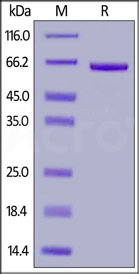
Biotinylated Human FGL1, Avitag,Fc Tag on SDS-PAGE under reducing (R) condition. The gel was stained with Coomassie Blue. The purity of the protein is greater than 95%.


活性(Bioactivity)-ELISA

FGL1 ELISA

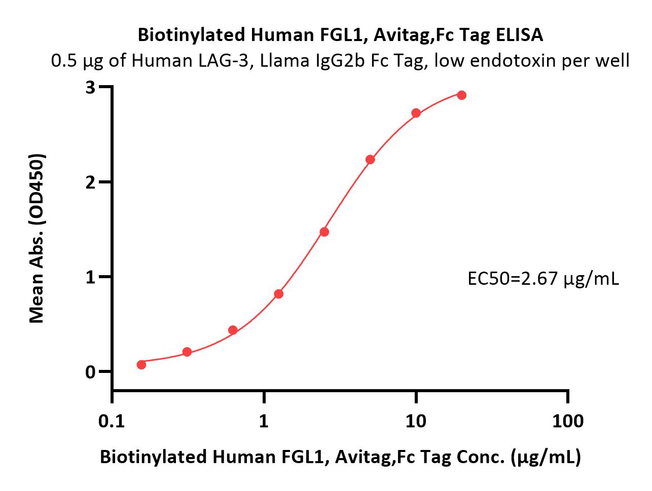
Immobilized Human LAG-3, Llama IgG2b Fc Tag, low endotoxin (Cat. No. LA3-H525c) at 5 μg/mL (100 μL/well) can bind Biotinylated Human FGL1, Avitag,Fc Tag (Cat. No. FG1-H82F4) with a linear range of 0.156-5 μg/mL (QC tested).

Protocol

FGL1 ELISA

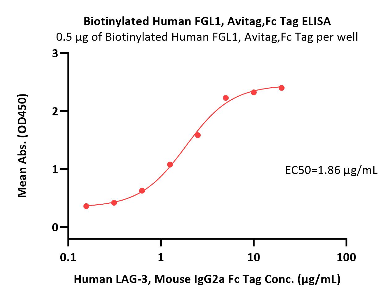
Immobilized Biotinylated Human FGL1, Avitag,Fc Tag (Cat. No. FG1-H82F4) at 5 μg/mL (100 μL/well)on streptavidin (Cat. No. STN-N5116) (2 μg/well) plate. can bind Human LAG-3, Mouse IgG2a Fc Tag (Cat. No. LA3-H52Aa) with a linear range of 0.156-2.5 μg/mL (Routinely tested).

Protocol


活性(Bioactivity)-BLI

FGL1 BLI

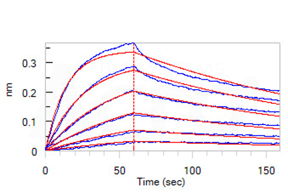
Loaded Biotinylated Human FGL1, Avitag,Fc Tag (Cat. No. FG1-H82F4) on SA Biosensor, can bind Human LAG-3, Fc Tag (Cat. No. LA3-H5255) with an affinity constant of 4.12 nM as determined in BLI assay (ForteBio Octet Red96e) (Routinely tested).

Protocol


活性(Bioactivity)-FACS

FGL1 FACS

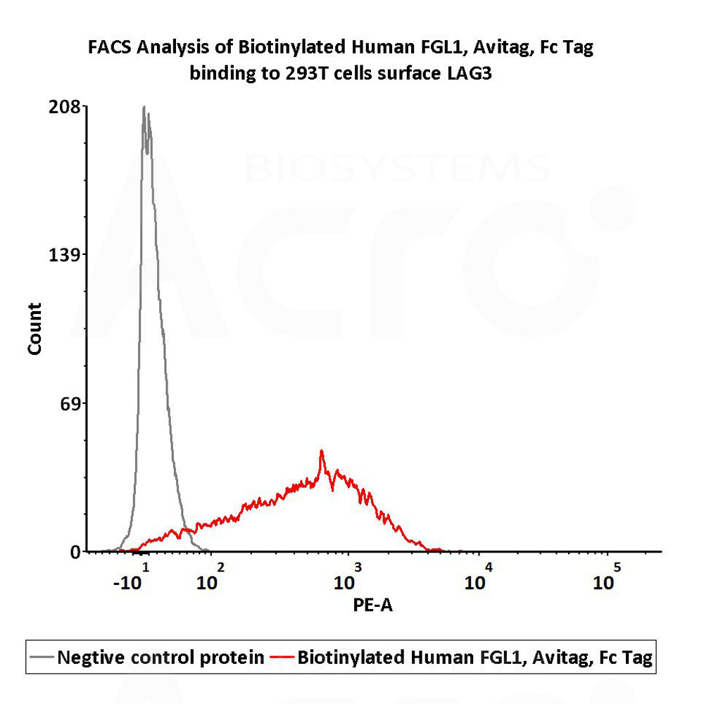
FACS analysis shows that Biotinylated Human FGL1, Avitag,Fc Tag (Cat. No. FG1-H82F4) can bind to 293T cells overexpressing human LAG3. The concentration of Human FGL1 is 10 μg/mL (Routinely tested).

Protocol

 
评论(0)
 
ACRO质量管理体系


背景(Background)

Fibrinogen-like protein 1(FGL1) is also known as HP-041, Hepassocin, HFREP-1, LFIRE-1. The protective effect of fibrinogen-like protein 1 (FGL1) in liver injury has previously been reported. However, studies have shown that FGL1 may be a predictor of GC patients and a target for GC therapy. Immunocytochemical studies revealed that fgl1 selectively binds to defective spermatozoa in the cauda epididymidis. Northern blot analysis and in situ hybridization demonstrated the high expression of fgl1 in the principal cells of the proximal cauda epididymidis. Immunofluorescence analysis using mouse fibrotic lung tissues suggested that fibrotic regions showed increased expressions of Gtse1 and Fgl1, Gtse1 and Fgl1 are suggested to be novel targets for radiation-induced lung fibrosis.


前沿进展

Monocyte-derived extracellular vesicles, stimulated by Trypanosoma cruzi, enhance cellular invasion in vitro via activated TGF-β1
Ansa-Addo, Pathak, McCrossan et al
J Extracell Vesicles (2024) 13 (11), e70014
Abstract: During cell invasion, large Extracellular Vesicle (lEV) release from host cells was dose-dependently triggered by Trypanosoma cruzi metacyclic trypomastigotes (Mtr). This lEV release was inhibited when IP3-mediated Ca2+ exit from the ER and further Ca2+ entry from plasma membrane channels was blocked, but whilst any store-independent Ca2+ entry (SICE) could continue unabated. That lEV release was equally inhibited if all entry from external sources was blocked by chelation of external Ca2+ points to the major contributor to Mtr-triggered host cell lEV release being IP3/store-mediated Ca2+ release, SICE playing a minor role. Host cell lEVs were released through Mtr interaction with host cell lipid raft domains, integrins, and mechanosensitive ion channels, whereupon [Ca2+]cyt increased (50 to 750 nM) within 15 s. lEV release and cell entry of T. cruzi, which increased up to 30 and 60 mpi, respectively, as well as raised actin depolymerization at 60 mpi, were all reduced by TRPC inhibitor, GsMTx-4. Vesicle release and infection was also reduced with RGD peptide, methyl-β-cyclodextrin, knockdown of calpain and with the calpain inhibitor, calpeptin. Restoration of lEV levels, whether with lEVs from infected or uninfected epithelial cells, did not restore invasion, but supplementation with lEVs from infected monocytes, did. We provide evidence of THP-1 monocyte-derived lEV interaction with Mtr (lipid mixing by R18-dequenching; flow cytometry showing transfer to Mtr of R18 from R18-lEVs and of LAP(TGF-β1). Active, mature TGF-β1 (at 175 pg/×105 in THP-1 lEVs) was detected in concentrated lEV-/cell-free supernatant by western blotting, only after THP-1 lEVs had interacted with Mtr. The TGF-β1 receptor (TβRI) inhibitor, SB-431542, reduced the enhanced cellular invasion due to monocyte-lEVs.© 2024 The Author(s). Journal of Extracellular Vesicles published by Wiley Periodicals LLC on behalf of International Society for Extracellular Vesicles.
ADAMTS16 drives epithelial-mesenchymal transition and metastasis through a feedback loop upon TGF-β1 activation in lung adenocarcinoma
Xiao, Li, Chen et al
Cell Death Dis (2024) 15 (11), 837
Abstract: Lung adenocarcinoma (LUAD) is the major subtype of lung cancer. The poor prognosis of LUAD patients is attributed primarily to metastasis. ADAMTS16 is a crucial member of the ADAMTS family and is involved in tumor progression. However, its role and regulatory mechanism in LUAD remain unexplored. In this study, ADAMTS16 was identified as a crucial oncogene and survival predictor in LUAD via analyses of public datasets. Clinical specimens and tissue microarrays confirmed the differential expression and prognostic value of ADAMTS16 in LUAD patients. Transcriptome data and in vitro experiments demonstrated that ADAMTS16 was positively associated with epithelial-mesenchymal transition (EMT) and the migration abilities of LUAD cells. Knockdown of ADAMTS16 attenuated lung and pleural metastasis in an animal model. Mechanistically, the results of the enzyme-linked immunosorbent assay (ELISA) and western blot (WB) suggested that ADAMTS16 activated the TGF-β signaling pathway by facilitating the conversion of LAP-TGF-β1 to active TGF-β1. Co-Immunoprecipitation (co-IP) indicated an interaction between ADAMTS16 and LAP-TGF-β1. Inhibition of ADAMTS16 impaired EMT and aggressiveness of LUAD cells, while treatment with recombinant TGF-β1 reversed this inhibition. Chromatin immunoprecipitation (ChIP) and dual-luciferase reporter assays indicated that SOX4 acted as a transcriptional activator of ADAMTS16 and that TGF-β1 regulated the expression of ADAMTS16 by increasing the binding of SOX4 to the promoter of ADAMTS16. Suppressing the TGF-β signaling pathway inhibited ADAMTS16 expression, EMT, and lung metastasis, whereas overexpressing SOX4 reversed this inhibition. Therefore, ADAMTS16 forms a positive feedback loop with the TGF-β1/SOX4 axis to regulate EMT and metastasis, and disruption of this feedback loop inhibits tumor progression. These findings underscore the potential of ADAMTS16 as a prognostic biomarker and therapeutic target in LUAD and offer novel insight into the mechanism of EMT and metastasis.© 2024. The Author(s).
Hypoxia Promotes the Expression of ADAM9 by Tubular Epithelial Cells, Which Enhances Transforming Growth Factor β1 Activation and Promotes Tissue Fibrosis in Patients With Lupus Nephritis
Umeda, Karino, Satyam et al
Arthritis Rheumatol (2025) 77 (2), 180-189
Abstract: Enhanced expression of transforming growth factor (TGF) β in the kidneys of patients with lupus nephritis (LN) can lead to progressive fibrosis, resulting in end-organ damage. ADAM9 activates TGFβ1 by cleaving the latency-associated peptide (LAP). We hypothesized that ADAM9 in the kidney may accelerate fibrogenesis by activating TGFβ1.We assessed the expression of ADAM9 in the kidneys of mice and humans who were lupus prone. In vitro experiments were conducted using tubular epithelial cells (TECs) isolated from mice and explored the mechanisms responsible for the up-regulation of ADAM9 and the subsequent activation of TGFβ1. To assess the role of ADAM9 in the development of tubular-intestinal fibrosis in individuals with LN, we generated MRL/lpr mice who were Adam9 deficient.ADAM9 was highly expressed in tubules from MRL/lpr mice. The transcription factor hypoxia-inducible factor-1α was found to promote the transcription of ADAM9 in TECs. TECs from mice who were Adam9 deficient and exposed to the hypoxia mimetic agent dimethyloxalylglycine failed to cleave the LAP to produce bioactive TGFβ1 from latent TGFβ1. Coculture of TECs from mice who were Adam9 deficient with fibroblasts in the presence of dimethyloxalylglycine and latent TGFβ1 produced decreased amounts of type I collagen and α-smooth muscle actin (SMA) by fibroblasts. MRL/lpr mice who were Adam9 deficient showed reduced interstitial fibrosis. At the translational level, ADAM9 expression in tissues and urine of patients with LN was found to increase.Hypoxia promotes the expression of ADAM9 by TECs, which is responsible for the development of interstitial fibrosis in patients with LN by enhancing the TGFβ1 activation, which promotes fibroblasts to produce collagen and α-SMA.© 2024 American College of Rheumatology.
Heterozygous mutations in the straitjacket region of the latency-associated peptide domain of TGFB2 cause Camurati-Engelmann disease type II
Wang, Kometani, Zeitlin et al
J Hum Genet (2024) 69 (11), 599-605
Abstract: Camurati-Engelmann disease (CED) is an autosomal dominant bone dysplasia characterized by progressive hyperostosis of the skull base and diaphyses of the long bones. CED is further divided into two subtypes, CED1 and CED2, according to the presence or absence of TGFB1 mutations, respectively. In this study, we used exome sequencing to investigate the genetic cause of CED2 in three pedigrees and identified two de novo heterozygous mutations in TGFB2 among the three patients. Both mutations were located in the region of the gene encoding the straitjacket subdomain of the latency-associated peptide (LAP) of pro-TGF-β2. Structural simulations of the mutant LAPs suggested that the mutations could cause significant conformational changes and lead to a reduction in TGF-β2 inactivation. An activity assay confirmed a significant increase in TGF-β2/SMAD signaling. In vitro osteogenic differentiation experiment using iPS cells from one of the CED2 patients showed significantly enhanced ossification, suggesting that the pathogenic mechanism of CED2 is increased activation of TGF-β2 by loss-of-function of the LAP. These results, in combination with the difference in hyperostosis patterns between CED1 and CED2, suggest distinct functions between TGFB1 and TGFB2 in human skeletal development and homeostasis.© 2024. The Author(s), under exclusive licence to The Japan Society of Human Genetics.
Showing 1-4 of 232 papers.
Powered by BizGenius
 
 
货号/价格
产品推荐
文档
联系电话:
+86 400-682-2521(全国)
010-53681107(北京)
021-50850665(上海)
运输方式
订单邮箱:
order.cn@acrobiosystems.com
技术支持邮箱:
tech.cn@acrobiosystems.com
FGL1靶点信息
英文全称:Fibrinogen-like protein 1
中文全称:
种类:
上市药物数量:0详情
临床药物数量:0详情
最高研发阶段:
查看更多信息
前沿进展
点击查看详细

消息提示

请输入您的联系方式,再点击提交!

确定