登录 | 注册    关注公众号  
微信公众号
搜索
 >  Protein>EGF R >EGR-HF2H5

FITC-Labeled Human EGF R Protein, His Tag DMF

分子别名(Synonym)

EGFR,ERBB,ERBB1,HER1,PIG61,mENA

表达区间及表达系统(Source)

FITC-Labeled Human EGF R, His Tag (EGR-HF2H5) is expressed from human 293 cells (HEK293). It contains AA Leu 25 - Ser 645 (Accession # P00533-1). It is the FITC labeled form of Human EGF R, His Tag (EGR-H5222).

Predicted N-terminus: Leu 25

Request for sequence

蛋白结构(Molecular Characterization)

EGF R Structure

This protein carries a polyhistidine tag at the C-terminus.

The protein has a calculated MW of 70.5 kDa. The protein migrates as 85-105 kDa under reducing (R) condition (SDS-PAGE) due to glycosylation.

偶联(Conjugate)

FITC

Excitation source: 488 nm spectral line, argon-ion laser

Excitation Wavelength: 488 nm

Emission Wavelength: 535 nm

标记(Labeling)

The primary amines in the side chains of lysine residues and the N-terminus of the protein are conjugated with FITC using standard chemical labeling method. The residual FITC is removed by molecular sieve treatment during purification process.

蛋白标记度(Protein Ratio)

The FITC to protein molar ratio is 2-3.5.

内毒素(Endotoxin)

Less than 1.0 EU per μg by the LAL method.

纯度(Purity)

>95% as determined by SDS-PAGE.

制剂(Formulation)

Lyophilized from 0.22 μm filtered solution in PBS, pH7.4 with trehalose as protectant.

Contact us for customized product form or formulation.

重构方法(Reconstitution)

Please see Certificate of Analysis for specific instructions.

For best performance, we strongly recommend you to follow the reconstitution protocol provided in the CoA.

存储(Storage)

For long term storage, the product should be stored at lyophilized state at -20°C or lower.

Please protect from light and avoid repeated freeze-thaw cycles.

This product is stable after storage at:

  1. -20°C to -70°C for 12 months in lyophilized state;
  2. -70°C for 3 months under sterile conditions after reconstitution.

质量管理控制体系(QMS)

  1. 质量管理体系(ISO, GMP)
  2. 质量优势
  3. 质控流程
 

电泳(SDS-PAGE)

EGF R SDS-PAGE

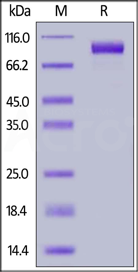
FITC-Labeled Human EGF R, His Tag on SDS-PAGE under reducing (R) condition. The gel was stained with Coomassie Blue. The purity of the protein is greater than 95%.


活性(Bioactivity)-ELISA

EGF R ELISA

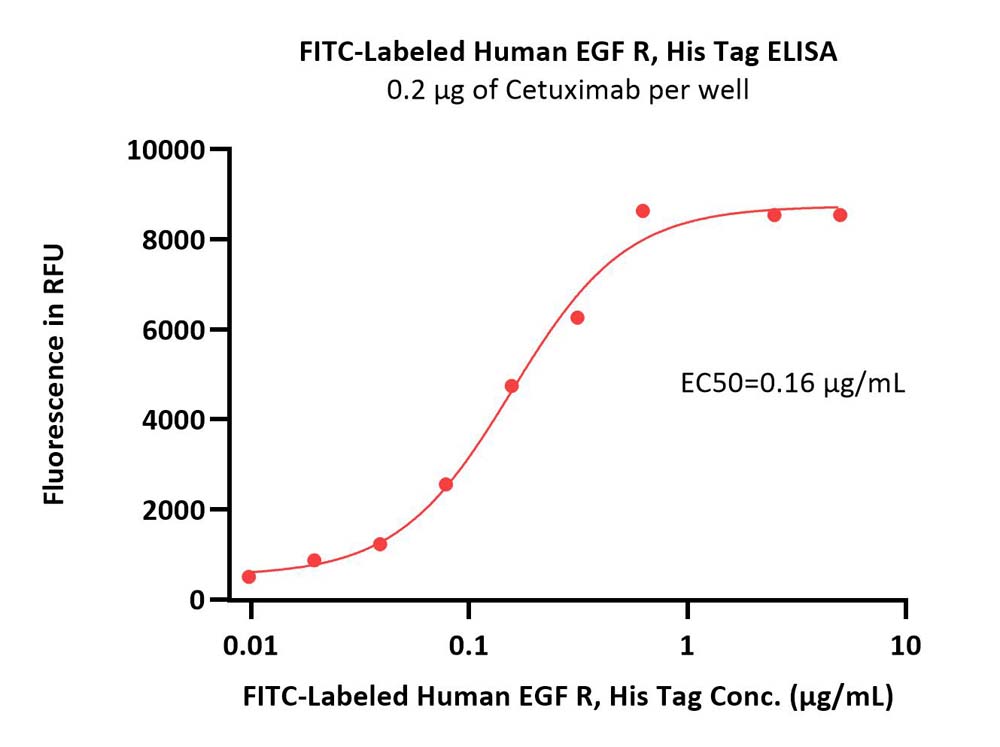
Immobilized Cetuximab at 2 μg/mL (100 μL/well) can bind FITC-Labeled Human EGF R, His Tag (Cat. No. EGR-HF2H5) with a linear range of 0.039-0.313 μg/mL (QC tested).

Protocol


活性(Bioactivity)-FACS

EGF R FACS

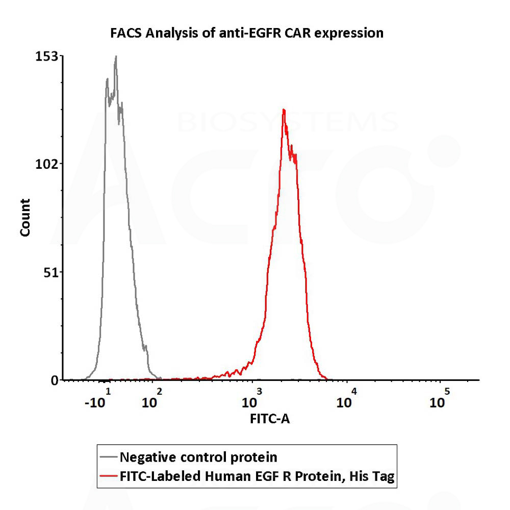
2e5 of Anti-EGFR CAR-293 cells were stained with 100 μL of 1 μg/mL of FITC-Labeled Human EGF R Protein, His Tag (Cat. No. EGR-HF2H5) and negative control protein respectively, FITC signal was used to evaluate the binding activity (QC tested).

Protocol

 
评论(0)
 
ACRO质量管理体系


背景(Background)

The epidermal growth factor receptor (EGFR; ErbB-1; HER1 in humans) is the cell-surface receptor for members of the epidermal growth factor family (EGF-family) of extracellular protein ligands. The epidermal growth factor receptor is a member of the ErbB family of receptors, a subfamily of four closely related receptor tyrosine kinases: EGFR (ErbB-1), HER2/c-neu (ErbB-2), Her 3 (ErbB-3) and Her 4 (ErbB-4). Mutations affecting EGFR expression or activity could result in cancer.

文献引用(Citations)


前沿进展

Global Bibliometric Visualization of Epidermal Growth Factor in Wound Healing: A Comprehensive Literature Search
Zyoud
Aesthetic Plast Surg (2025)
Abstract: Epidermal growth factor (EGF) is essential for both wound repair and the maintenance of healthy tissues. Numerous bibliometric studies have been published on wound healing; however, none have focused on EGF in wound healing. This study comprehensively analyzed scientific publications on the role of Epidermal Growth Factor (EGF) in wound healing. The analysis encompassed publications indexed in the Scopus database from 1972 to 2024, retrieved using a broad range of relevant keywords. Network analysis and visualization were subsequently conducted using VOSviewer (v. 1.6.20) to map research trends and identify research hotspots. My examination of the Scopus database identified 3122 articles on the use of EGF in wound healing published over the past 48 years (1972-2024). Most of these publications were research articles (n = 2812; 90.07%), followed by review articles (n = 244; 7.82%). The number of publications on the use of EGF in wound healing has increased significantly (R2=0.9449; p < 0.001) over the years. This study revealed that, during the past ten years, there has been an increasing amount of global research on the relationship between EGF and wound healing. The effectiveness and clinical applications of human EGF and EGFR signaling in cancer-related wound healing were the main topics of our study, which focused on EGF and wound healing overall. To improve patient outcomes and enhance the efficacy of wound healing, particularly in aesthetic and reconstructive surgery, this bibliometric analysis emphasizes the importance of ongoing research and collaboration in this field.Level of Evidence V This journal requires that authors assign a level of evidence to each article. For a full description of these Evidence-Based Medicine ratings, please refer to the Table of Contents or the online Instructions to Authors www.springer.com/00266 .© 2025. Springer Science+Business Media, LLC, part of Springer Nature and International Society of Aesthetic Plastic Surgery.
Revealing the multi-target compounds of Sarcandra glabra identification and inhibition of novel target genes for the treatment of pancreatic cancer
Liu, Ou
BMC Complement Med Ther (2025) 25 (1), 106
Abstract: S. glabra has been widely used to treat tumors in traditional Chinese medicine (TCM). However, the specific mechanism of action of S. glabra in pancreatic cancer remains unclear. In this study, network pharmacological analysis was used to identify the active components of S. glabra and their corresponding targets for the treatment of pancreatic cancer. Furthermore, molecular docking, molecular dynamic simulations, and in vitro experiments were performed to validate the findings.The active components of S. glabra and their corresponding targets for the treatment of pancreatic cancer were identified using the TCMSP database and a literature search. Differentially expressed genes were identified using data from the Gene Expression Omnibus (GEO) database, and their protein-protein interaction (PPI) network was constructed using the STRING platform. The target genes of S. glabra were further assessed using Gene Ontology (GO) and Kyoto Encyclopedia of Genes and Genomes (KEGG) analyses in the R software. Subsequently, a protein-protein interaction (PPI) network and a composite target-pathway network were established. The target genes were subjected to survival and mutation analyses. Molecular docking and molecular dynamic simulations were used to validate the interaction between the hub target genes and S. glabra in vitro. In addition, cell viability and qRT-PCR verification of S. glabra against pancreatic cancer in vitro.A total of 20 active components and 70 targets were identified. Based on the PPI network, CASP3, MMP9, CCND1, EGF, MMP2, CASP8, ERBB2, STAT1, and PPARG were identified as hub target genes. Enrichment analysis showed that S. glabra may primarily affect pathways such as p53 signaling, transcriptional dysregulation in cancer, proteoglycans in cancer, pancreatic cancer, and cell cycle. Molecular docking and molecular dynamic simulations indicated stable binding between anhydroicaritin-GSK3B and quercetin-PPARG. In vitro experiments demonstrated that treatment with S. glabra significantly inhibited the growth of PANC-1 cells and downregulated expression of GSK3B and PPARG (P < 0.05).This study demonstrates the potential of S. glabra, a herb in traditional Chinese medicine, for treating pancreatic cancer. The findings provide insights into the mechanism of action of the active ingredients of S. glabra, offering a strong theoretical foundation for its various clinical applications.Not applicable.© 2025. The Author(s).
snRNA-Seq and Spatial Transcriptome Reveal Cell-Cell Crosstalk Mediated Metabolic Regulation in Porcine Skeletal Muscle
Guo, Han, Xu et al
J Cachexia Sarcopenia Muscle (2025) 16 (2), e13752
Abstract: Cell-cell crosstalk between myogenic, adipogenic and immune cells in skeletal muscle to regulate energy metabolism and lipid deposition has received considerable attention. The specific mechanisms of interaction between the different cells in skeletal muscle are still unclear.Using integrated analysis of snRNA-seq and spatial transcriptome, the gene expression profile of longissimus dorsi (LD) muscle was compared between adult Taoyuan black (TB, obese, native Chinese breed) and Duroc (lean) pigs.TB pig had more intramuscular fat (IMF) deposition (3.91%, p = 0.0244) and higher slow myofiber proportion (17.13%, p < 0.0001) compared with Duroc pig (IMF, 2.38%; slow myofiber, 6.92%) at the age of 180 days. We identified eight cell populations in porcine LD muscle. Five subpopulations of myonuclei and 10 subclusters of fibro/adipogenic progenitors (FAPs) were defined by marker genes. CellChat analysis revealed that communication between immune cells and other cells via the BMP and EGF signalling pathway was only observed in Duroc and not in TB pig. Both snRNA-seq and spatial transcriptome pointed out that FAPs are the important source of secretory proteins. A total of 35 upregulated and 23 downregulated differentially expressed genes (DEGs) were annotated as secretory, one upregulated and 36 downregulated secretory DEGs were identified between TB and Duroc pigs in FAPs by snRNA-seq and FAPs-high regions by spatial transcriptome, respectively. The distribution of FAPs was accompanied by the divergent myofiber-type composition. The expression level of slow myofiber marker gene (MYH7) was higher in both FAPs-high and FAPs-low regions of TB compared with Duroc pig (p < 0.0001), and expression level of fast myofiber maker gene (MYH1) was upregulated in FAPs-high region of Duroc compared with FAPs-high region of TB (p < 0.0001) and FAPs-low region of Duroc pig (p = 0.0002). The metabolic differences of myofibers between TB and Duroc pigs were mainly concentrated in energy, lipid and nitrogen metabolism-related pathway (p < 0.05). The significant correlation (R > 0.4, p < 0.05) between secretory and metabolism-related DEGs with spatial aggregation was verified by regression analysis for random region extraction (area of 25 spots, n = 400) from spatial transcriptome, and we speculated that the alteration of secretory proteins forming the microenvironment might regulate myofiber metabolism via target genes such as IRS1, PLPP1 and SLC38A2.Our study provides new insights into skeletal muscle microenvironment that contributes to metabolic regulation and new methods and resources to study cell-cell communication in skeletal muscle.© 2025 The Author(s). Journal of Cachexia, Sarcopenia and Muscle published by Wiley Periodicals LLC.
Adhesion-Assisted Antioxidant-Engineered Mesenchymal Stromal Cells for Enhanced Cardiac Repair in Myocardial Infarction
Zhong, Luo, Wu et al
ACS Nano (2025)
Abstract: Mesenchymal stromal cell (MSC) therapy holds great promise for treating myocardial infarction (MI). However, the inflammatory and reactive oxygen species (ROS)-rich environment in infarcted myocardium challenges MSC survival, limiting its therapeutic impact. In this study, we demonstrate that chemical modification of MSCs with anti-VCAM1 and polydopamine (PD) significantly enhances MSC survival and promotes cardiac repair. Anti-VCAM1 modification facilitates MSC adhesion to inflamed tissue, ensuring MSC retention in the injured myocardium, while PD scavenges ROS surrounding MSCs, creating a conducive environment for cell transplantation. Our data indicate that chemically engineered MSCs effectively disrupt the inflammation-ROS cycle and modulate inflammation-related immune responses, thus improving MI microenvironments. Single-cell RNA sequencing of rat hearts reveals that treatment with engineered MSCs inhibits cardiac fibrosis by suppressing HB-EGF-EGFR signaling between anti-inflammatory macrophages and activated fibrillates. Ultimately, engineered MSCs demonstrate superior therapeutic efficacy in a rat model of MI. This study presents a straightforward, safe, and efficient chemical method for enhancing MSC therapy.
Showing 1-4 of 3245 papers.
Powered by BizGenius
 
 
货号/价格
文档
联系电话:
+86 400-682-2521(全国)
010-53681107(北京)
021-50850665(上海)
运输方式
订单邮箱:
order.cn@acrobiosystems.com
技术支持邮箱:
tech.cn@acrobiosystems.com
EGF R靶点信息
英文全称:Epidermal growth factor receptor erbB1
中文全称:表皮生长因子受体erbB1
种类:Homo sapiens
上市药物数量:33详情
临床药物数量:182详情
最高研发阶段:批准上市
查看更多信息
前沿进展
点击查看详细

消息提示

请输入您的联系方式,再点击提交!

确定