Whole genome sequencing of 378 prostate cancer metastases reveals tissue selectivity for mismatch deficiency with potential therapeutic implicationsVis, Palit, Corradi
et alGenome Med (2025) 17 (1), 24
Abstract: Survival of patients with metastatic castration-resistant prostate cancer (mCRPC) depends on the site of metastatic dissemination.Patients with mCRPC were prospectively included in the CPCT-02 metastatic site biopsy study. We evaluated whole genome sequencing (WGS) of 378 mCRPC metastases to understand the genetic traits that affect metastatic site distribution.Our findings revealed that RB1, PIK3CA, JAK1, RNF43, and TP53 mutations are the most frequent genetic determinants associated with site selectivity for metastatic outgrowth. Furthermore, we explored mutations in the non-coding genome and found that androgen receptor (AR) chromatin binding sites implicated in metastatic prostate cancer differ in mutation frequencies between metastatic sites, converging on pathways that impact DNA repair. Notably, liver and visceral metastases have a higher tumor mutational load (TML) than bone and lymph node metastases, independent of genetic traits associated with neuroendocrine differentiation. We found that TML is strongly associated with DNA mismatch repair (MMR)-deficiency features in these organs.Our results revealed gene mutations that are significantly associated with metastatic site selectivity and that frequencies of non-coding mutations at AR chromatin binding sites differ between metastatic sites. Immunotherapeutics are thus far unsuccessful in unselected mCRPC patients. We found a higher TML in liver and visceral metastases compared to bone and lymph node metastases. As immunotherapeutics response is associated with mutational burden, these findings may assist in selecting mCRPC patients for immunotherapy treatment based on organs affected by metastatic disease.NCT01855477.© 2025. The Author(s).
Genomic alterations in the WNT/β-catenin pathway and resistance of colorectal cancer cells to pathway-targeting therapiesVoutsadakis
Explor Target Antitumor Ther (2025) 6, 1002295
Abstract: Colorectal cancer is the most prevalent gastrointestinal malignancy with limited therapeutic options in the metastatic setting. The WNT/β-catenin/adenomatous polyposis coli (APC) pathway is commonly deregulated in the disease and presents a rational target for therapeutic exploitation.The publicly available genomic data from the colorectal cancer cohort of the Cancer Genome Atlas (TCGA) were used to define groups of colorectal cancers with alterations in APC or other key genes of the WNT/β-catenin/APC pathway and to identify genomic characteristics of interest in each group. In vitro sensitivity data for drugs targeting the pathway were compiled from the Genomics of Drug Sensitivity in Cancer (GDSC) project.Three-fourths of colorectal cancers possessed APC alterations and about one in four of these cases possessed also concomitant alterations in other genes of the WNT/β-catenin/APC pathway, including RNF43, CTNNB1, and TCF7L2. Colorectal cancers with alterations in one or more of the three genes of the WNT/β-catenin pathway, RNF43, CTNNB1, and TCF7L2, in the absence of APC alterations, were frequently microsatellite instability (MSI) high and had high tumor mutation burden (TMB). Cancers with these same alterations in the three genes with or without APC alterations presented a high frequency of mutations in receptor tyrosine kinases, PI3K/AKT pathway genes, and DNA damage response genes. Cell lines without mutations in WNT/β-catenin/APC pathway components displayed numerically greater sensitivity to inhibitors of the pathway in vitro.Groups of colorectal cancers differing in WNT/β-catenin/APC pathway alterations present diverse genomic landscapes that could have therapeutic implications for the rational development of inhibitors of the pathway.© The Author(s) 2025.
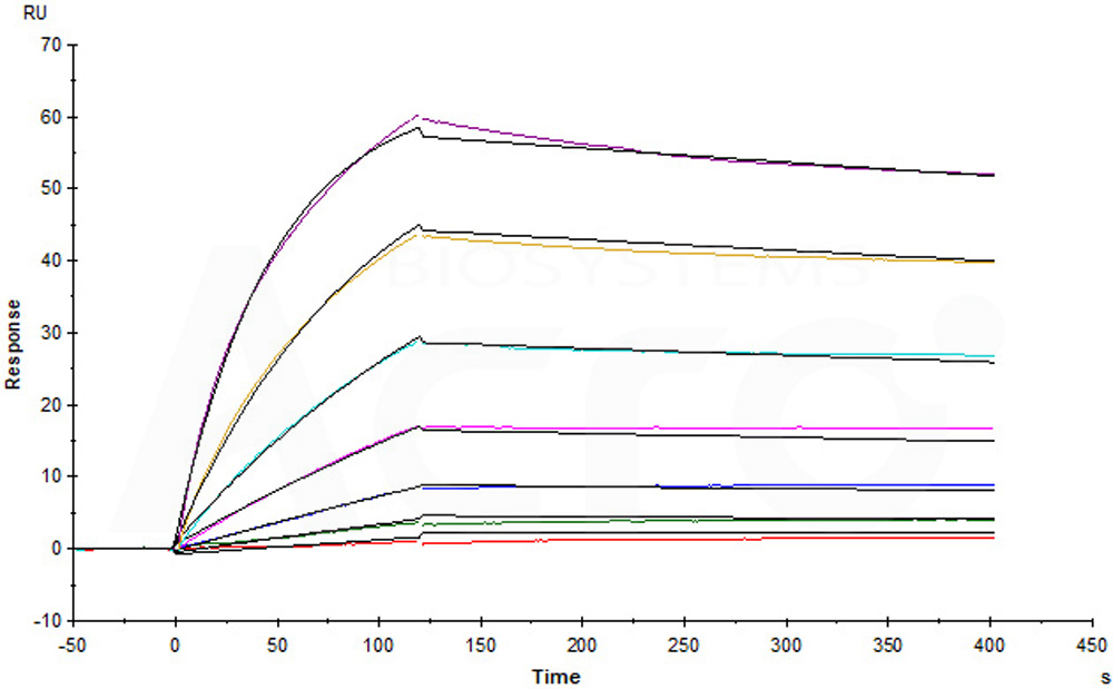
Structure-Guided Development of Chemically Tailored Peptide Binders of RNF43/ZNRF3 to Enable Versatile Design of Membrane Protein-Targeting PROTACsCui, Zheng, Weng
et alAngew Chem Int Ed Engl (2025)
Abstract: Targeted membrane protein degradation using cell surface E3 ligases RNF43/ZNRF3 via proteolysis targeting chimeras (PROTACs) represents an effective strategy for treating membrane drug targets that cannot be fully inhibited using traditional inhibitors. Several ingenious chimeras have been developed to tether RNF43/ZNRF3 to target membrane proteins, resulting in the degradation of targets at sub-nanomolar concentrations both in vitro and in vivo. However, currently available RNF43/ZNRF3 binders are genetically encoded and have poor plasticity, which limits the design and promotion of such PROTACs. Here, we exploited the AlphaFold-predicted complex structures of ligand-bound RNF43/ZNRF3 and developed a class of chemically tailored peptide binders for ZNRF3/RNF43. With these peptide binders that can be conveniently prepared by de novo peptide synthesis, we established a new membrane protein degradation platform that allows versatile modular design and targeted degradation of clinically relevant membrane proteins, i.e., PD-L1 and EGFR. This study presents a new subtype within the PROTAC field to develop therapeutic peptides targeting membrane proteins.© 2025 Wiley‐VCH GmbH.
Assessing Genotype-Phenotype Correlations with Deep Learning in Colorectal Cancer: A Multi-Centric StudyGustav, van Treeck, Reitsam
et almedRxiv (2025)
Abstract: Deep Learning (DL) has emerged as a powerful tool to predict genetic biomarkers directly from digitized Hematoxylin and Eosin (H&E) slides in colorectal cancer (CRC). However, few studies have systematically investigated the predictability of biomarkers beyond routinely available alterations such as microsatellite instability (MSI), and BRAF and KRAS mutations.Our primary dataset comprised H&E slides of CRC tumors across five cohorts totaling 1,376 patients who underwent comprehensive panel sequencing, with an additional 536 patients from two public datasets for validation. We developed a DL model using a single transformer model to predict multiple genetic alterations directly from the slides. The model's performance was compared against conventional single-target models, and potential confounders were analyzed.The multi-target model was able to predict numerous biomarkers from pathology slides, matching and partly exceeding single-target transformers. The Area Under the Receiver Operating Characteristic curve (AUROC, mean ± std) on the primary external validation cohorts was: BRAF (0·78 ± 0·01), hypermutation (0·88 ± 0·01), MSI (0·93 ± 0·01), RNF43 (0·86 ± 0·01); this biomarker predictability was mirrored across metrics and co-occurrence analyses. However, biomarkers with high AUROCs largely correlated with MSI, with model predictions depending considerably on MSI-associated morphology upon pathological examination.Our study demonstrates that multi-target transformers can predict the biomarker status for numerous genetic alterations in CRC directly from H&E slides. However, their predictability is mainly associated with MSI phenotype, despite indications of slight biomarker-inherent contributions to a phenotype. Our findings underscore the need to analyze confounders in AI-based oncology biomarkers. To enable this, we developed a validated model applicable to other cancers and larger, diverse datasets.The German Federal Ministry of Health, the Max-Eder-Programme of German Cancer Aid, the German Federal Ministry of Education and Research, the German Academic Exchange Service, and the EU.


 膜杰作
膜杰作 Star Staining