登录 | 注册    关注公众号  
微信公众号
搜索
 >  Beads>Spike protein >MBS-K031

SARS-CoV-2 Spike Trimer (P.1) Coupled Magnetic Beads

热销产品推荐:The Magnetic Stand (Cat.No. MB-01 & Cat.No. MB-02) can be used in conjunction with Beads.

ItemsSize (2mg)Size(5mg X 2)
Particle size2 μm2 μm
Physical appearancePowder mixturePowder mixture
Amount of Coupled Protein≈286 pmol (40 μg) SARS-CoV-2 Spike Trimer (P.1)/mg beads≈286 pmol (40 μg) SARS-CoV-2 Spike Trimer (P.1)/mg beads
Binding Capacity>242 pmol (36 μg) Anti-SARS-CoV-2 Spike RBD Neutralizing Antibody/mg beads>242 pmol (36 μg) Anti-SARS-CoV-2 Spike RBD Neutralizing Antibody/mg beads
FormulationPBS, pH7.4, with 10% TrehalosePBS, pH7.4, with 10% Trehalose
Reconstitution2 mL sterile deionized water (1 mg beads/mL)5 mL sterile deionized water (1 mg beads/mL)

背景(Background)

The SARS-CoV-2 Spike Trimer (P.1) Coupled Magnetic Beads is produced by coupling biotinylated SARS-CoV-2 spike trimer to streptavidin-conjugated magnetic beads. The spike proteins coupled to the beads contain 12 mutations (L18F, T20N, P26S, D138Y, R190S, K417T, E484K, N501Y, D614G, H655Y, T1027I, V1176F) identified in the Brazilian variant (known as P.1). The pre-coupled beads are ready to use for capturing anti-SARS-CoV-2 antibody or ACE2 protein from your sample with high specificity.

表达区间及表达系统(Source)

SARS-CoV-2 Spike Trimer (P.1) Protein is expressed from human 293 cells (HEK293). It contains AA Val 16 - Pro 1213(Accession # QHD43416.1 (L18F, T20N, P26S, D138Y, R190S, K417T, E484K, N501Y, D614G, H655Y, T1027I, V1176F, R683A, R685A,F817P, A892P, A899P, A942P, K986P, V987P)).

应用说明(Application)

This product is intended for immunocapture, biopanning. This is a non-sterile product.

*The Isotype control (Cat. No. SMB-B01) is sold separately.

重构方法(Reconstitution)

See Certificate of Analysis (CoA) for detailed instruction.

存储(Storage)

Upon receipt, please store the Beads at -20°C. The shelf life is 1 year at -20 °C.

Please avoid more than 3 freeze-thaw cycles once reconstitution, immediate use after reconstitution is highly recommended.

原理(Assay Principles)

Antibody Purification: 1. Resuspend the lyophilized beads by adding the buffer of choice. 2. Add analyte to the suspension, mix and incubate to enable specific binding of the beads and the target protein. 3. Magnetize beads, remove supernatant, and wash unbound protein fractions to capture target protein-bound beads. 4. Wash, magnetize the beads and collect purified target protein for use in downstream applications.

The magnetic beads technology makes use of the easy and efficient collection of beads in magnetic field to facilitate antibody purification in a simple workflow of “bind-wash-elute”. In contrast to common separation techniques, this method does not require columns or centrifugation, and is therefore ideal in high-throughput applications.

制剂(Formulation)

Please contact us for detailed information.

Contact us for customized product form or formulation.

质量管理控制体系(QMS)

  1. 质量管理体系(ISO, GMP)
  2. 质量优势
  3. 质控流程
 

典型数据-Typical Data

Spike protein TYPICAL DATA

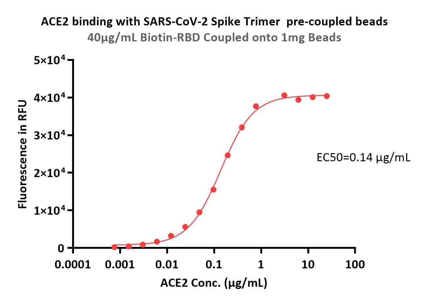
Immobilized 40 μg SARS-CoV-2 S protein trimer to 1mg Beads can bind the ACE2 with an EC50 of 0.14μg/mL (QC tested).

Protocol

Spike protein TYPICAL DATA

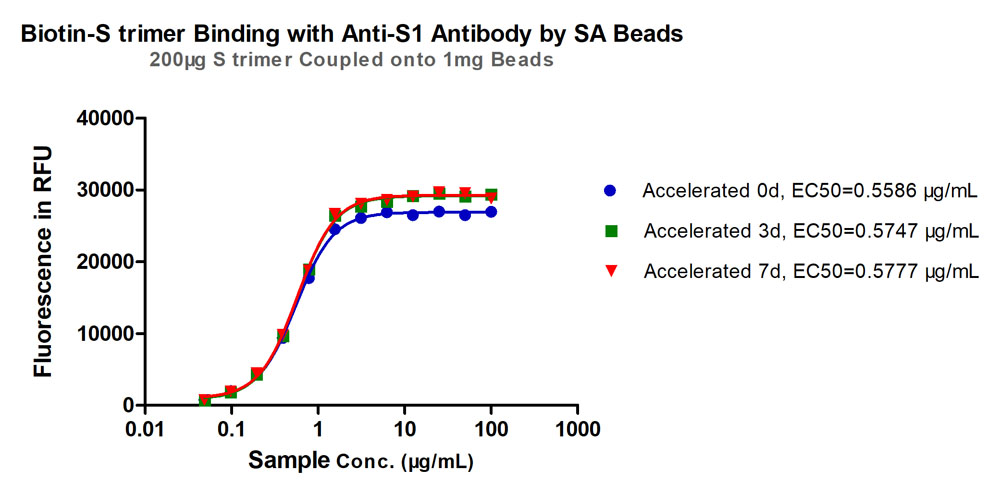
Accelerated stability test. After placing the lyophilized beads at 37°C for 7 days, binding activity between the S SARS-CoV-2 Spike Trimer (P.1) Coupled Magnetic Beads (Cat.No. MBS-K031) and anti-SARS-CoV-2 Spike S1 antibody showed little deviation from the unaccelerated sample (%RSD<5%). Data were measured on day 0, 3, 7 respectively.

Protocol

Spike protein TYPICAL DATA

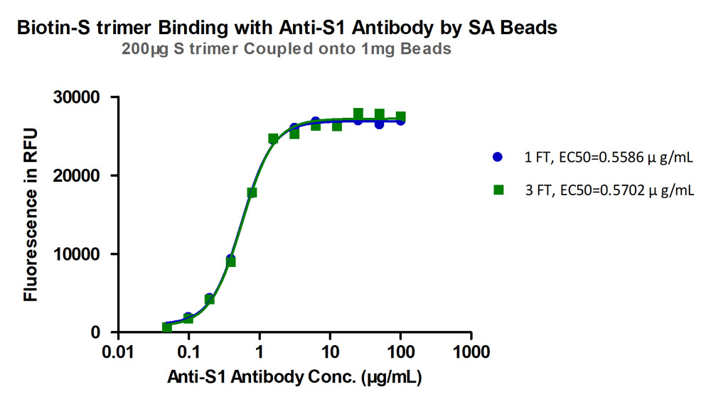
Freeze-thaw stability test. After different freeze-thaw cycles, binding activity between SARS-CoV-2 Spike Trimer (P.1) Coupled Magnetic Beads (Cat.No. MBS-K031) and anti-SARS-CoV-2 Spike S1 antibody showed little deviation from the unfree-thaw sample (%RSD<5%). Three freeze-thaw cycles were performed.

Protocol

 
评论(0)
 
ACRO质量管理体系


前沿进展

The XBB.1.5 mRNA booster vaccine does not significantly increase the percentage of XBB.1.5 mono-reactive T cells
Sop, Mercado, Figueroa et al
Front Immunol (2025) 16, 1513175
Abstract: Recent efforts in vaccine development have targeted spike proteins from evolving SARS-CoV-2 variants. In this study, we analyzed T cell responses to the XBB.1.5 and BA.2.86 subvariants in individuals who previously received bivalent vaccines containing mRNA for ancestral and BA.5 spike proteins. T cell-mediated cytokine responses to spike proteins from both variants were largely preserved. To determine the mechanism of this preserved recognition, we utilized the functional expansion of specific T cells (FEST) assay to distinguish between the presence of T cells that cross-recognized ancestral and variant epitopes versus distinct populations of T cells that were mono-reactive for ancestral or variant epitopes. We found the majority of spike-specific T cells cross-recognized the ancestral spike and the XBB.1.5 and BA.2.86 subvariants, with less than 10% of T cells being mono-reactive for either variant. Interestingly, immunization with the XBB.1.5 monovalent booster vaccine did not significantly increase the percentage of XBB.1.5 mono-reactive T cells. Our results suggest a potential limitation in the induction of mono-reactive T cell responses by variant-specific booster vaccines.Copyright © 2025 Sop, Mercado, Figueroa, Beckey, Traut, Zhang, Smith and Blankson.
Antibody levels and the risk of SARS-CoV-2 infection during the Omicron surge
Sasaki, Kadowaki, Matsumoto et al
GHM Open (2024) 4 (1), 52-53
Abstract: We examined the association between antibody titer levels and risk of coronavirus disease 2019 (COVID-19) infection in the general Japanese population, including a total of 1,972 participants between June and September 2022. Specifically, we ascertained participantsIgG antibody titers targeting the spike protein and infection status, and subsequently examined the association between antibody titer categories (< 2,500, 2,500-5,000, 5,000-10,000 and > 10,000 AU/mL) and COVID-19 infection to estimate risk ratios (RR) and their 95% confidence intervals (CI). Compared to the lowest category, the adjusted RR for participants with antibody titers ≥ 10,000 AU/mL was 0.38 (95% CI: 0.20-0.71). The observed non-linear relationship between the titers and the risk of infection showed that the risk decreased as the participant's antibody titer increased, but the slope became milder when the antibody titer reached approximately 10,000 AU/mL. These findings may contribute to the use of an individual's antibody titer to consider appropriate timing of vaccination.2024, National Center for Global Health and Medicine.
Long-term antibody response after COVID-19 vaccination in health care workers: A single centre study from Pakistan
Kanani, Ejaz, Anis et al
J Pak Med Assoc (2025) 75 (3), 472-475
Abstract: The retrospective cohort study was planned to determine long-term anti-spike immunoglobulin G levels after receiving coronavirus disease-2019 vaccination by healthcare workers. The study took place between June and July 2022 at the Indus hospital in Karachi. Healthcare workers who had previously screened negative to prevaccination Severe Acute Respiratory Syndrome Coronavirus 2 nucleocapsid antibodies were tested for post-vaccination Severe Acute Respiratory Syndrome Coronavirus 2 anti-spike immunoglobulin G levels using a quantitative assay. The test was also performed on the stored pre-vaccination samples of the subjects collected up to 18 months previously. Antibody levels in subjects without infection, with infection and with booster administration were compared. The median postvaccination anti-spike immunoglobulin G in infected only, infected with routine vaccination and infected with booster values were 1,725.6 IU/mL (interquartile range: 684.80- 4,708.9 IU/mL), 2,067.15 IU/mL (interquartile range: 705.33-4,670.4 IU/mL) and 6,139.15 IU/mL (interquartile range: 2,426.05-10,623.40 IU/mL). There was a doubling of antibody titers, from 1,744 IU/mL to 3,829 IU/mL, in those who received a booster versus routine vaccination (p>0.05). The antibodies remained positive more than a year following vaccination.
Within-Host Fitness and Antigenicity Shift Are Key Factors Influencing the Prevalence of Within-Host Variations in the SARS-CoV-2 S Gene
Xi, Hua, Jiang et al
Viruses (2025) 17 (3)
Abstract: Within-host evolution plays a critical role in shaping the diversity of SARS-CoV-2. However, understanding the primary factors contributing to the prevalence of intra-host single nucleotide variants (iSNVs) in the viral population remains elusive. Here, we conducted a comprehensive analysis of over 556,000 SARS-CoV-2 sequencing data and prevalence data of different SARS-CoV-2 S protein amino acid mutations to elucidate key factors influencing the prevalence of iSNVs in the SARS-CoV-2 S gene. Within-host diversity analysis revealed the presence of mutational hotspots within the S gene, mainly located in NTD, RBD, TM, and CT domains. Additionally, we generated a single amino acid resolution selection status map of the S protein. We observed a significant variance in within-host fitness among iSNVs in the S protein. The majority of iSNVs exhibited low to no within-host fitness and displayed low alternate allele frequency (AAF), suggesting that they will be eliminated due to the narrow transmission bottleneck of SARS-CoV-2. Notably, iSNVs with moderate AAFs (0.06-0.12) were found to be more prevalent than those with high AAFs. Furthermore, iSNVs with the potential to alter antigenicity were more prevalent. These findings underscore the significance of within-host fitness and antigenicity shift as two key factors influencing the prevalence of iSNVs in the SARS-CoV-2 S gene.
Showing 1-4 of 39008 papers.
Powered by BizGenius
 
 
货号/价格
文档
联系电话:
+86 400-682-2521(全国)
010-53681107(北京)
021-50850665(上海)
运输方式
订单邮箱:
order.cn@acrobiosystems.com
技术支持邮箱:
tech.cn@acrobiosystems.com
Spike protein靶点信息
英文全称:Coronavirus spike glycoprotein
中文全称:冠状病毒刺突糖蛋白
种类:Severe acute respiratory syndrome coronavirus 2 (2019-nCoV) (SARS-CoV-2)
上市药物数量:10详情
临床药物数量:72详情
最高研发阶段:批准上市
查看更多信息
前沿进展
点击查看详细

消息提示

请输入您的联系方式,再点击提交!

确定