登录 | 注册    关注公众号  
微信公众号
搜索
 >  Protein>TSPAN8 >TS8-H52H3

Human TSPAN8 Protein, His Tag

热销产品推荐:

ACROBiosystems提供针对多种生物标志物靶标的lHC抗体产品,具有:验证充分、质量可靠、灵活授权及免疫组化检测服务支持!

分子别名(Synonym)

CO-029,tetraspanin 8,TM4SF3,TM4SF3tspan-8,Transmembrane 4 superfamily member 3tetraspanin-8,TSPAN8,Tspan-8,Tumor-associated antigen CO-029

表达区间及表达系统(Source)

Human TSPAN8, His Tag (TS8-H52H3) is expressed from human 293 cells (HEK293). It contains AA Lys 110 - Asn 205 (Accession # P19075-1).

Predicted N-terminus: His

Request for sequence

蛋白结构(Molecular Characterization)

TSPAN8 Structure

This protein carries a polyhistidine tag at the N-terminus.

The protein has a calculated MW of 12.9 kDa. The protein migrates as 14 kDa and 17-19 kDa under reducing (R) condition (SDS-PAGE) due to glycosylation.

内毒素(Endotoxin)

Less than 1.0 EU per μg by the LAL method.

纯度(Purity)

>90% as determined by SDS-PAGE.

制剂(Formulation)

Lyophilized from 0.22 μm filtered solution in PBS, pH7.4 with trehalose as protectant.

Contact us for customized product form or formulation.

重构方法(Reconstitution)

Please see Certificate of Analysis for specific instructions.

For best performance, we strongly recommend you to follow the reconstitution protocol provided in the CoA.

存储(Storage)

For long term storage, the product should be stored at lyophilized state at -20°C or lower.

Please avoid repeated freeze-thaw cycles.

This product is stable after storage at:

  1. -20°C to -70°C for 12 months in lyophilized state;
  2. -70°C for 3 months under sterile conditions after reconstitution.

质量管理控制体系(QMS)

  1. 质量管理体系(ISO, GMP)
  2. 质量优势
  3. 质控流程
 

电泳(SDS-PAGE)

TSPAN8 SDS-PAGE

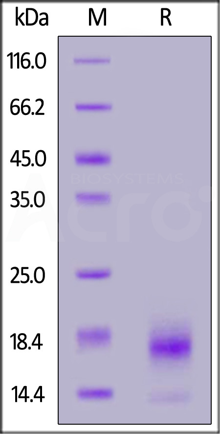
Human TSPAN8, His Tag on SDS-PAGE under reducing (R) condition. The gel was stained with Coomassie Blue. The purity of the protein is greater than 90%.


活性(Bioactivity)-ELISA

TSPAN8 ELISA

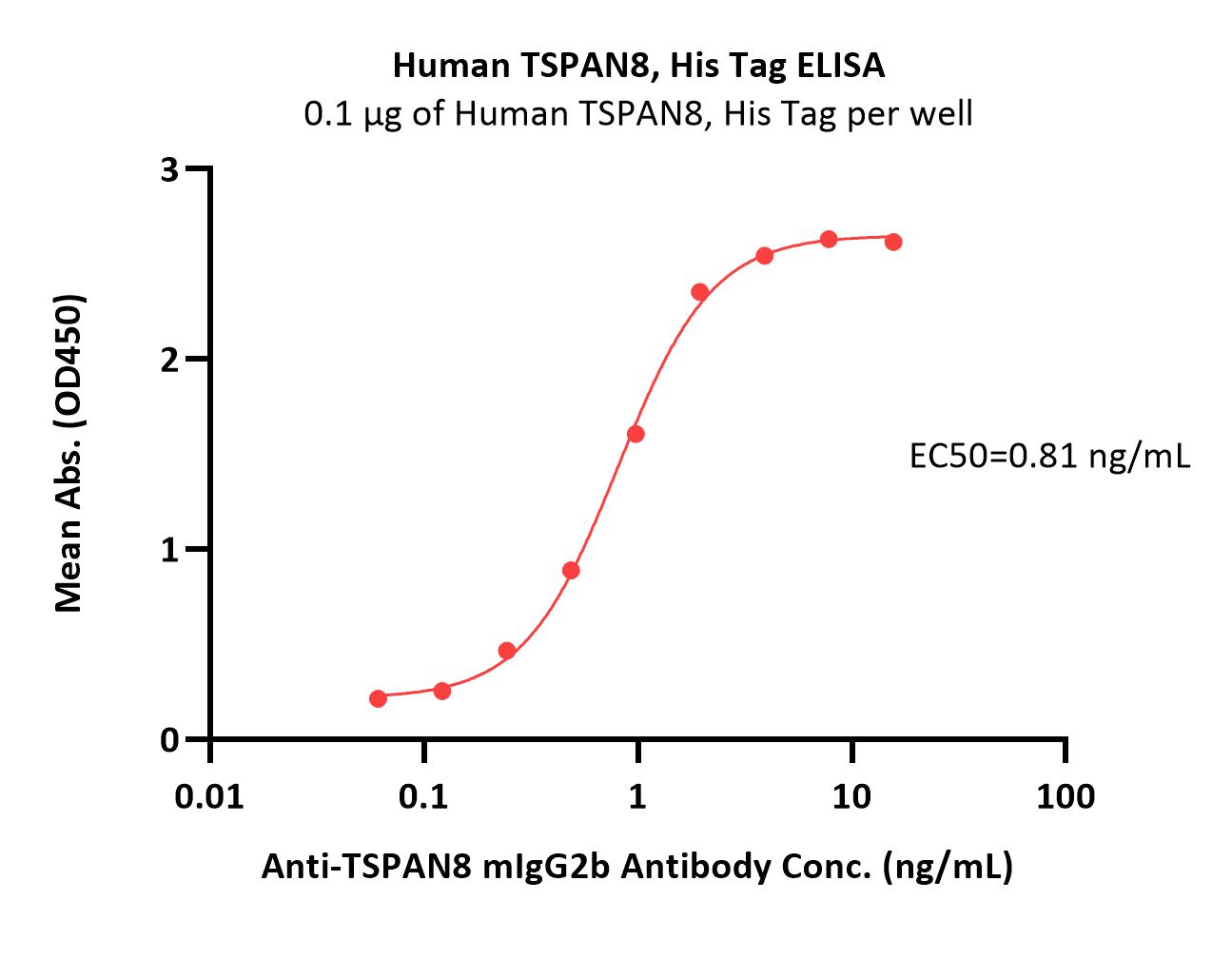
Immobilized Human TSPAN8, His Tag (Cat. No. TS8-H52H3) at 1 μg/mL (100 μL/well) can bind Anti-TSPAN8 mIgG2b Antibody with a linear range of 0.06-2 ng/mL (QC tested).

Protocol

TSPAN8 ELISA

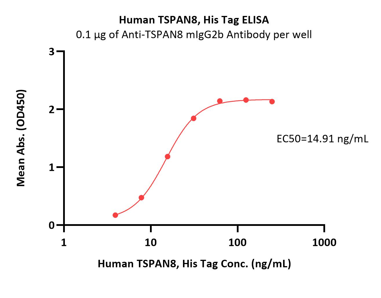
Immobilized Anti-TSPAN8 mIgG2b Antibody at 1 μg/mL (100 μL/well) can bind Human TSPAN8, His Tag (Cat. No. TS8-H52H3) with a linear range of 4-63 ng/mL (Routinely tested).

Protocol

 
评论(1)
  1. 159XXXXXXX6
  2. 0人赞
  3. 我们使用此蛋白主要用于检测抗体的结合活性,从ELISA实验的结果可以看出:能较好的拟合成一条“S”型曲线。蛋白稳定,复溶效果好。
  4. 2024-8-24
 
ACRO质量管理体系


背景(Background)

Tspan8 is 1 of the 33 mammalian members of the tetraspanin family, composed of transmembrane proteins that organize laterally, together or with other membrane partners such as integrins, to form ‘tetraspanin webs’. These platforms signal within cells to regulate many cellular processes: adhesion, migration, invasion or survival Tspan8 has been implicated in many types of cancer.Overexpression was reported in glioma and colorectal, esophageal, hepatic, gastric and pancreatic carcinoma. Tspan8 exerts a pro-invasive function by controlling cell–cell and cell–matrix interactions through its association with membrane partners such as α6β4 integrin-protein kinase C (PKC)-activated, E-cadherin, EpCAM, claudin-7 and CD44. Moreover, Tspan8 may be a promising new therapeutic target, as Tspan8-specific antibodies were shown to reduce cell motility, block tumor angiogenesis in vivo.


前沿进展

Characterization of LINC00483 as a Novel Oncogene in Colorectal Cancer via Targeting miR-601/TSPAN8 Signaling Axis
Liu, Xiong, Chen et al
J Biochem Mol Toxicol (2025) 39 (3), e70164
Abstract: LINC00483 is exerted a crucial function in many malignant tumors' progression, but the role of LINC00483 in colorectal cancer (CRC) carcinogenesis remains not fully understood. The aim of our study was to explore the potential role of LINC00483 in the development of CRC and its underlying molecular mechanism. In the present study, the level of mRNA and protein was assessed through using qRT-PCR and Western blot analysis assays. Cell proliferation was assessed by using Cell Counting Kit-8 (CCK-8) and colony formation assays. The cell metastasis was assessed by using transwell assays. Results revealed that LINC00483 was elevated in CRC tissue and cell lines. Upregulation of LINC00483 was negatively correlated with the overall survival. The mRNA expression of tetraspanin 8 (TSPAN8) is increased in CRC tissues, whereas the expression of miR-601 is decreased, and in CRC tissues, a correlation between the expression levels of LINC00483 and TSPAN8 was positive, but the correlation between LINC00483 and miR-601 was negative. LINC00483 deficiency could inhibit the cell proliferation, migration, and invasion in CRC cell lines. Moreover, the proliferation, migration, and invasion were inhibited in CRC cell lines after transfected with miR-601 mimics; but this effect could be eliminated by LINC00483 overexpressed. Results also suggested that the protein expression of TSPAN8 and N-cadherin is decreased in CRC cell lines after transfected with miR-601 mimics, whereas the protein expression of E-cadherin is increased; but this effect could be abrogated by LINC00483 overexpressed, which means that miR-601 mimics can inhibit the metastasis of CRC, but after cotransfection with LINC00483, the metastasis ability of CRC is restored. In conclusion, our study elucidated that LINC00483 promote the CRC progression by upregulating TSPAN8 via sponging miR-601. This finding provided a new potential drug target for the treatment of CRC in clinic.© 2025 Wiley Periodicals LLC.
Single cell RNA-sequencing of Ewing sarcoma tumors demonstrates transcriptional heterogeneity and clonal evolution
Goodspeed, Bodlak, Duffy et al
Clin Cancer Res (2025)
Abstract: Ewing sarcoma (EwS) is the second most common bone cancer in children, accounting for 2% of pediatric cancer diagnoses. Patients who present with metastatic disease at the time of diagnosis have a dismal prognosis, compared to the >70% 5-year survival of those with localized disease. Novel therapeutic approaches that can impact metastatic disease are desperately needed, as well as a deeper understanding of the heterogeneity of EwS tumors.Here, we utilized single cell RNA-sequencing to characterize the transcriptional landscape of primary EwS tumors and surrounding tumor microenvironment (TME) in a cohort of seven untreated EwS patients, as well as in circulating tumor cells (CTCs). A potential CTC therapeutic target was evaluated through immunofluorescence of fixed CTCs from a separate cohort.Primary tumor samples demonstrate a heterogenous transcriptional landscape with several conserved gene expression programs, including those composed of genes related to proliferation and EWS gene targets, which were found to correlate with overall survival. Copy-number analysis identified subclonal evolution within patients prior to treatment. Analyses of the immune microenvironment reveal an immunosuppressive microenvironment with complex intercellular communication within the tumor and immune cells. Single cell RNA-sequencing and immunofluorescence of CTCs at the time of diagnosis identified TSPAN8 as a potential therapeutic target.EwS tumors demonstrate significant transcriptional heterogeneity as well as a complex immunosuppressive microenvironment. This work evaluates several proposed targets which would warrant further exploration as novel therapeutic strategies.
[Transcriptomic differences between the spleens of mice immunized with inactivated antigens of foot-and-mouth disease virus and Senecavirus A]
Zheng, Ma, Li et al
Sheng Wu Gong Cheng Xue Bao (2024) 40 (12), 4493-4508
Abstract: The aim of this study was to compare the immune responses of C57BL/6 mice immunized with two pathogens, foot-and-mouth disease virus (FMDV) and Senecavirus A (SVA), and to provide clues for revealing the regulatory mechanisms of acquired immunity. Inactivated and purified FMDV and SVA antigens were used to immunize C57BL/6 mice respectively, and the mice immunized with PBS were taken as the control. The percentages of Th1 and Th2 cells in the spleen lymphocytes of mice in each group were analyzed by flow cytometry at 14 and 28 days after immunization. RNA-Seq was performed for the spleen. Mouse macrophages were stimulated with the antigens in vitro to examine the expression of the differentially expressed genes (DEGs) screened out. The results showed that 14 days after immunization, there was no significant difference in the magnitude of the Th1/Th2 immune response elicited by the FMDV and SVA antigens. After 28 days, the magnitudes of the Th1 and Th2 immune responses elicited by the SVA antigen were higher than those elicited by the FMDV antigen. RNA-Seq revealed two common DEGs, Rsad2 and Tspan8, between the two immunization groups, which indicated that the two genes may be involved in the activation of the Th1/Th2 immune responses by FMDV and SVA antigens. FMDV and SVA antigens stimulated macrophages to secrete interleukin (IL)-12 and IL-33 in vitro, and the expression of Tspan8 and Rsad2 was consistent with the RNA-Seq results. The expression of Rsad2 was regulated by type I interferons (IFNα, IFNβ). In this study, we obtained the DEGs involved in the immune responses to the two antigens in mouse spleen, which provides a molecular basis for investigating the immune response mechanisms induced by FMDV and SVA.
Showing 1-4 of 228 papers.
Powered by BizGenius
 
 
货号/价格
文档
联系电话:
+86 400-682-2521(全国)
010-53681107(北京)
021-50850665(上海)
运输方式
订单邮箱:
order.cn@acrobiosystems.com
技术支持邮箱:
tech.cn@acrobiosystems.com
TSPAN8靶点信息
英文全称:Tetraspanin-8
中文全称:四跨膜蛋白-8
种类:
上市药物数量:0详情
临床药物数量:0详情
最高研发阶段:终止
查看更多信息
前沿进展
点击查看详细

消息提示

请输入您的联系方式,再点击提交!

确定