登录 | 注册    关注公众号  
微信公众号
搜索
 >  Protein>CD98 >CD8-HF248

FITC-Labeled Human CD98 Protein, His Tag

分子别名(Synonym)

SLC3A2,CD98,4F2,4F2HC,4T2HC,CD98HC,MDU1,NACAE

表达区间及表达系统(Source)

FITC-Labeled Human CD98, His Tag (CD8-HF248) is expressed from human 293 cells (HEK293). It contains AA Arg 206 - Ala 630 (Accession # P08195-1).

Predicted N-terminus: His

Request for sequence

蛋白结构(Molecular Characterization)

Online(Arg 206 - Ala 630) P08195-1

This protein carries a polyhistidine tag at the N-terminus.

The protein has a calculated MW of 47.7 kDa. The protein migrates as 55-66 kDa under reducing (R) condition (SDS-PAGE) due to glycosylation.

Conjugate

FITC

Excitation source: 488 nm spectral line, argon-ion laser

Excitation Wavelength: 488 nm

Emission Wavelength: 535 nm

标记(Labeling)

The primary amines in the side chains of lysine residues and the N-terminus of the protein are conjugated with FITC using standard chemical labeling method. The residual FITC is removed by molecular sieve treatment during purification process.

蛋白标记度(FITC:Protein Ratio)

The FITC to protein molar ratio is 1-3.

内毒素(Endotoxin)

Less than 1.0 EU per μg by the LAL method.

纯度(Purity)

>95% as determined by SDS-PAGE.

制剂(Formulation)

Lyophilized from 0.22 μm filtered solution in PBS, pH7.4. Normally trehalose is added as protectant before lyophilization.

Contact us for customized product form or formulation.

重构方法(Reconstitution)

Please see Certificate of Analysis for specific instructions.

For best performance, we strongly recommend you to follow the reconstitution protocol provided in the CoA.

存储(Storage)

For long term storage, the product should be stored at lyophilized state at -20°C or lower.

Please protect from light and avoid repeated freeze-thaw cycles.

This product is stable after storage at:

  1. -20°C to -70°C for 12 months in lyophilized state;
  2. -70°C for 3 months under sterile conditions after reconstitution.

质量管理控制体系(QMS)

  1. 质量管理体系(ISO, GMP)
  2. 质量优势
  3. 质控流程
 

电泳(SDS-PAGE)

FITC-Labeled Human CD98, His Tag (Cat. No. CD8-HF248) SDS-PAGE gel

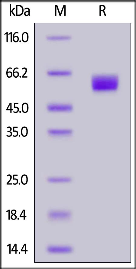
FITC-Labeled Human CD98, His Tag on SDS-PAGE under reducing (R) condition. The gel was stained with Coomassie Blue. The purity of the protein is greater than 95%.

 
评论(0)
 
ACRO质量管理体系


背景(Background)

CD antigen CD98 is also known as 4F2 cell-surface antigen heavy chain (4F2hc), 4F2 heavy chain antigen, Solute carrier family 3 member 2 (SLC3A2), Lymphocyte activation antigen 4F2 large subunit, is a single-pass type I I membrane protein which belongs to the SLC3A transporter family.. SLC3A2 / CD98 is expressed ubiquitously in all tissues tested with highest levels detected in kidney, placenta and testis and weakest level in thymus. SLC3A2 / CD98 is required for the function of light chain amino-acid transporters and also involved in sodium-independent, high-affinity transport of large neutral amino acids such as phenylalanine, tyrosine, leucine, arginine and tryptophan. CD98 involved in guiding and targeting of LAT1 and LAT2 to the plasma membrane. When associated with SLC7A5 or SLC7A8, CD98 involved in the cellular activity of small molecular weight nitrosothiols, via the stereoselective transport of L-nitrosocysteine (L-CNSO) across the transmembrane. Together with ICAM1, regulates the transport activity LAT2 in polarized intestinal cells, by generating and delivering intracellular signals.


前沿进展

TBK1 is a signaling hub in coordinating stress-adaptive mechanisms in head and neck cancer progression
Kim, Kim, Kim et al
Autophagy (2025)
Abstract: Tumorigenesis is closely linked to the ability of cancer cells to activate stress-adaptive mechanisms in response to various cellular stressors. Stress granules (SGs) play a crucial role in promoting cancer cell survival, invasion, and treatment resistance, and influence tumor immune escape by protecting essential mRNAs involved in cell metabolism, signaling, and stress responses. TBK1 (TANK binding kinase 1) functions in antiviral innate immunity, cell survival, and proliferation in both the tumor microenvironment and tumor cells. Here, we report that MUL1 loss results in the hyperactivation of TBK1 in both HNC cells and tissues. Mechanistically, under proteotoxic stress induced by proteasomal inhibition, HSP90 inhibition, or Ub+ stress, MUL1 promotes the degradation of active TBK1 through K48-linked ubiquitination at lysine 584. Furthermore, TBK1 facilitates autophagosome-lysosome fusion and phosphorylates SQSTM1, regulating selective macroautophagic/autophagic clearance in HNC cells. TBK1 is required for SG formation and cellular protection. Moreover, we found that MAP1LC3B is partially localized within SGs. TBK1 depletion enhances the sensitivity of HNC cells to cisplatin-induced cell death. GSK8612, a novel TBK1 inhibitor, significantly inhibits HNC tumorigenesis in xenografts. In summary, our study reveals that TBK1 facilitates the rapid removal of ubiquitinated proteins within the cell through protective autophagy under stress conditions and assists SG formation through the use of the autophagy machinery. These findings highlight the potential of TBK1 as a therapeutic target in HNC treatment.Abbreviations: ALP: autophagy-lysosomal pathway; AMBRA1: autophagy and beclin 1 regulator 1; BaF: bafilomycin A1; CC: coiled-coil; CD274/PDL-1: CD274 molecule; CHX: cycloheximide; CQ: chloroquine; DNP: dinitrophenol; EGFR: epidermal growth factor receptor; ESCC: esophageal squamous cell carcinoma; G3BP1: G3BP stress granule assembly factor 1; HNC: head and neck cancer; HPV: human papillomavirus; IFN: interferon; IGFBP3: insulin like growth factor binding protein 3; IRF: interferon-regulatory factor 3; KO: knockout; LAMP1: lysosomal associated membrane protein 1; MAP1LC3B: microtubule associated protein 1 light chain 3 beta; NPC: nasopharyngeal carcinoma; PABP: poly(A) binding protein; PI: proteasome inhibitor; PQC: protein quality control; PROTAC: proteolysis-targeting chimera; PURA/PURα: purine rich element binding protein A; RIGI: RNA sensor RIG-I; SD: standard deviation; SG: stress granule; SQSTM1: sequestosome 1; STING1: stimulator of interferon response cGAMP interactor 1; TBK1: TANK binding kinase 1; UPS: ubiquitin-proteasome system; USP10: ubiquitin specific peptidase 10; VCP: valosin containing protein; VHL: von Hippel-Lindau tumor suppressor; WT: wild type.
Structural basis for the substrate recognition and transport mechanism of the human y+LAT1-4F2hc transporter complex
Dai, Zeng, Zhang et al
Sci Adv (2025) 11 (12), eadq0558
Abstract: Heteromeric amino acid transporters (HATs), including y+LAT1-4F2hc complex, are responsible for transporting amino acids across membranes, and mutations in y+LAT1 cause lysinuric protein intolerance (LPI), a hereditary disorder characterized by defective cationic amino acid transport. The relationship between LPI and specific mutations in y+LAT1 has yet to be fully understood. In this study, we characterized the function of y+LAT1-4F2hc complex in mammalian cells and determined the cryo-EM structures of the human y+LAT1-4F2hc complex in two distinct conformations: the apo state in an inward-open conformation and the native substrate-bound state in an outward-open conformation. Structural analysis suggests that Asp243 in y+LAT1 plays a crucial role in coordination with sodium ion and substrate selectivity. Molecular dynamic (MD) simulations further revealed the different transport mechanism of cationic amino acids and neutral amino acids. These results provide important insights into the mechanisms of the substrate binding and working cycle of HATs.
Suppression of NF-κB and downstream XBP1 by DcR3 contributes to a decrease in antibody secretion
Liu, Huang, Lin et al
J Immunol (2025) 214 (1), 72-84
Abstract: Decoy receptor 3 (DcR3), a soluble receptor in the tumor necrosis factor receptor superfamily, regulates the functions of monocytes, macrophages, dendritic cells, and T cells. Previous studies have demonstrated that DcR3 suppresses B cell proliferation in vitro and ameliorates autoimmune diseases in animal models; however, whether and how DcR3 regulates antibody production is unclear. Using a DcR3 transgenic mouse model, we found that DcR3 impaired the T cell-dependent antigen-stimulated antibody response. The number of Ag-specific antibody-secreting cells was transiently reduced, but the concentration of specific antibodies continued to decrease in the DcR3 transgenic mice, implying a direct suppression of antibody production by DcR3. In vitro assays showed that the DcR3-Fc fusion protein attenuated T cell-dependent induced antibody production and reduced the expression of secretory Igh and Xbp1. We found that nuclear factor κB (NF-κB) activity was essential for the expression of Xbp1 in activated B cells. DcR3-Fc attenuated anti-CD40-induced NF-κB activity and Xbp1 promoter activity. Furthermore, DcR3-Fc decreased the expression of Xbp1 in Blimp1+ antibody-secreting cells. Restoration of spliced XBP1 (X-box binding protein 1) in DcR3-treated B cells increased the secretory Ighg1 transcript levels, suggesting that reducing XBP1 is one of the mechanisms by which DcR3 regulates antibody production both in vitro and in vivo. Collectively, these results indicate that in addition to blocking proliferation, DcR3 impairs NF-κB activation, subsequently decreasing the expression of Xbp1, eventually leading to a reduction in antibody secretion.© The Author(s) 2025. Published by Oxford University Press on behalf of The American Association of Immunologists. All rights reserved. For commercial re-use, please contact reprints@oup.com for reprints and translation rights for reprints. All other permissions can be obtained through our RightsLink service via the Permissions link on the article page on our site—for further information please contact journals.permissions@oup.com.
Comparative Radiotracing Quantifies Brain Cellular Uptake and Catabolism of Bispecific Antibodies Targeting Transferrin Receptor and CD98hc
Veire, Lucas, Bond et al
ACS Chem Neurosci (2025)
Abstract: Bispecific antibodies (bAbs) that engage cerebrovascular targets, induce transport across the blood-brain barrier (BBB), and redistribute to secondary targets within the brain parenchyma have the potential to transform the diagnosis and treatment of a wide range of central nervous system disorders. Full understanding of the pharmacokinetics (PK) of these agents, including their potential for delivering cargo into brain parenchymal cells, is a key priority for the development of numerous potential therapeutic applications. To date, the brain PK of bAbs that target transferrin receptor (TfR-1) and CD98 heavy chain (CD98hc) has been characterized using techniques incapable of distinguishing between CNS clearance of intact protein from uptake and catabolism by brain parenchymal cells. Herein, we address this knowledge gap via a comparative radiotracing strategy using two radioisotopes with distinct residualizing properties, iodine-125 (I-125) and zirconium-89 (Zr-89). We first identify reaction conditions for tetravalent chelator modification and Zr-89 radiolabeling that do not adversely affect in vitro or in vivo function. We then use comparative radiotracing to define the PK of TfR-1 and CD98hc targeted bAbs without a parenchymal target, generating quantitative evidence of TfR-1-mediated cellular uptake and catabolism that implicates these processes in previously reported differences in the brain retention of IgGs shuttled across the BBB via these two pathways. Finally, we perform comparative radiotracing on a TfR-1 bAb with an internalizing neuronal target (TrkB), demonstrating rapid divergence of Zr-89 and I-125 PK curves, with a > 30-fold difference in brain content of the two radioisotopes. Together, these results establish comparative radiotracing as a valuable technique for identifying internalizing cellular targets within the brain parenchyma and quantifying the extent and timing of bAb uptake and catabolism following target engagement.
Showing 1-4 of 1988 papers.
Powered by BizGenius
 
 
货号/价格
文档
联系电话:
+86 400-682-2521(全国)
010-53681107(北京)
021-50850665(上海)
运输方式
订单邮箱:
order.cn@acrobiosystems.com
技术支持邮箱:
tech.cn@acrobiosystems.com
CD98靶点信息
英文全称:L-type amino acid transporter 1
中文全称:L-型氨基酸转运蛋白-1
种类:Homo sapiens
上市药物数量:0详情
临床药物数量:7详情
最高研发阶段:申请上市
查看更多信息
前沿进展
点击查看详细

消息提示

请输入您的联系方式,再点击提交!

确定