登录 | 注册    关注公众号  
微信公众号
搜索
 >  Kits>IL-4 >CRS-A004

resDetect™ Human Interleukin-4 (IL-4) ELISA Kit (Residue Testing)

For research use only.

产品参数(Product Specifications)

Assay TypeSandwich-ELISA
AnalyteIL-4
Format96T(8×12 strips)
ReactivityHuman
Regulatory StatusRUO
Sensitivity<0.391pg/mL
Standard Curve Range0.391 pg/mL-25 pg/mL
Assay Time2 hr 50 min
Suitable Sample TypeFor the quantitative determination of human IL-4 in Cell Culture Supernatants, Plasma, Serum.
Sample volume100 uL

产品概述(Product Overview)

resDetect™ Human Interleukin-4 (IL-4) ELISA Kit (Residue Testing) is based on the ELISA sandwich method and is used to detect and quantitatively determine GMP human IL-4 in cell culture supernatants, serum, and plasma. The kit contains GMP human IL-4 (ACROBiosystems, cat#GMP-L04H26) to ensure accurate assay results and is designed to provide a reliable solution for CAR-T product quality assessment during drug development and CMC quality control stages. It can also be used as a universal detection tool for the quantitative determination of human IL-4.

重构方法(Reconstitution)

Please see Certificate of Analysis for details of reconstitution instruction and specific concentration.

存储(Storage)

1. Unopened kit should be stored at 2℃-8℃ upon receiving.

2. Find the expiration date on the outside packaging and do not use reagents past their expiration date.

3. The opened kit should be stored per components table. The shelf life is 30 days from the date of opening.

组分(Materials Provided)

IDComponentsSize
CRS004-C01Pre-coated Anti-IL-4 Antibody Microplate1 plate(8×12 strips)
CRS004-C02Human IL-4 Standard20 μg
CRS004-C03Biotin-Anti-IL-4 Antibody50 μL
CRS004-C04Streptavidin-HRP50 μL
CRS004-C0510xWashing Buffer 50 mL
CRS004-C062xDilution Buffer50 mL
CRS004-C07Substrate Solution12 mL
CRS004-C08Stop Solution7 mL

质量管理控制体系(QMS)

  1. 质量管理体系(ISO, GMP)
  2. 质量优势
  3. 质控流程
 

典型数据-Typical Data Please refer to DS document for the assay protocol.

IL-4 TYPICAL DATA

For each experiment, a standard curve needs to be set for each micro-plate, and the specific OD value may vary depending on different laboratories, testers, or equipments. The following example data is for reference only.


验证(Validation)

稀释线性(Dilution Linearity)

To assess the linearity of the assay, samples spiked with high concentrations of human IL-4 were serially diluted with calibrator diluent to produce samples with values within the dynamic range of the assay.

IL-4 DILUTION LINEARITY

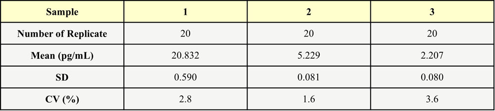
批内差异(Intra-Assay Statistics)

Three samples of known concentration were tested twenty times on one plate to assess intra-assay precision, Intra-Assay Precision CV<10%.

IL-4 INTRA-ASSAY STATISTICS

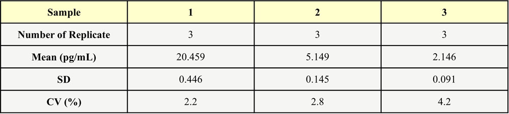
批间差异(Inter-Assay Statistics)

Three samples of known concentration were tested in three separate assays to assess inter-assay precision, Inter-Assay Precision CV<10%.
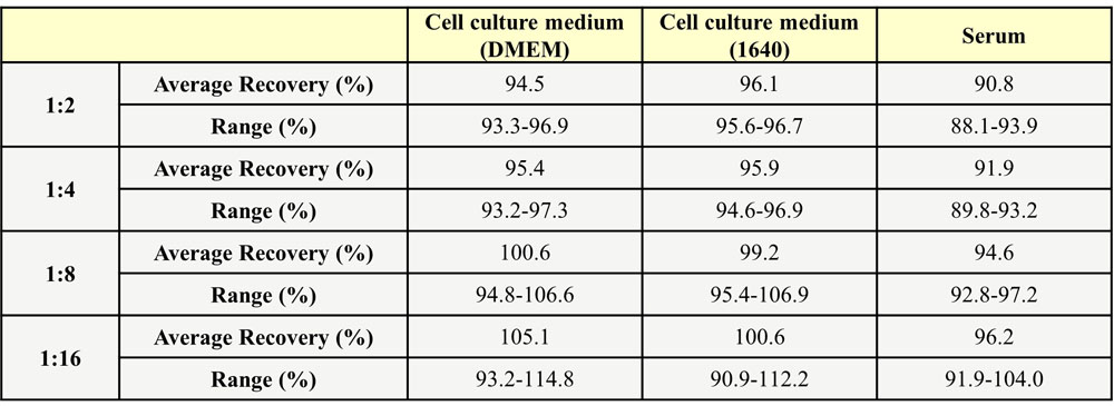
IL-4 INTER-ASSAY STATISTICS

回收率(Recovery)

Three parts of blank serum were added with different concentrations of human IL-4, and the serum without human IL-4 was used as background to calculate the recovery rate. The range of the recovery rate is 80.9-97.7%, and the average recovery is 86.3%.

IL-4 RECOVERY

 
评论(0)
 
ACRO质量管理体系


背景(Background):IL-4

Interleukin-4, is a cytokine that induces differentiation of naive helper T cells (Th0 cells to Th2 cells). In the presence of IL-4 and IL-13, cytokines that are produced in a Th-2 type response, particularly during allergy and parasitic infections, macrophages become differentially activated, And this cytokine is a ligand for interleukin 4 receptor. The interleukin 4 receptor also binds to IL13, which may contribute to many overlapping functions of this cytokine and IL13. STAT6, a signal transducer and activator of transcription, has been shown to play a central role in mediating the immune regulatory signal of this cytokine. Recently, researcher found that the cytokine IL-4 plays a key role in development of innate CD8+ T cells in the thymus of several gene-deficient mouse strains, including Itk, KLF2, CBP and Id3, without previous exposure to antigen.


前沿进展

Five-month real-ambient PM2.5 exposure impairs learning in Brown Norway rats: Insights from multi omics-based analysis
Song, Zhou, Xiong et al
Ecotoxicol Environ Saf (2025) 294, 118065
Abstract: PM2.5, recognized as a potential pathogenic factor for nervous system diseases, remains an area with many unknowns, particularly regarding its effects on human health. After five-month real-ambient PM2.5 exposure, we observed no significant pathological damage to the lung, liver, spleen, or kidney tissues. However, PM2.5 exposure led to neuronal degeneration in the hippocampal CA1 region of Brown Norway (BN) rats. The level of IL-6, IL-13, IL-1β, IL-12, IL-4, GRO/KC, MIP-1α, CM-CSF significantly increased in lung lavage fluid (P < 0.05 for all). Notably, we detected a slight impairment in spatial learning ability, as evidenced by the Barnes maze training outcomes. There were no significant changes in the bacterial community in lung lavage fluid (P = 0.621), but the bacterial community in the gut significantly changed (P < 0.001), with more species identified (P < 0.05). The metabolomic analysis revealed 147 and 149 significantly changed metabolites in the pulmonary system and serum, respectively (P < 0.05). PM2.5 exposure caused a decrease in Nervonic acid (NA) in both the lung and serum, which likely contributed to spatial learning impairment (P < 0.01). The correlation between lung metabolites, gut bacterial species, and serum metabolites indicated that PM2.5 exposure likely impaired spatial learning through the lung-gut-brain axis pathway. Lung and serum metabolic disorders and intestinal microbial imbalance occurred in BN rats post-five-month real-ambient PM2.5 exposure. There were two potential ways that PM2.5 exposure caused the decline of spatial learning ability in wild-type BN rats: (1) PM2.5 exposure led to a significant decrease of neuroprotective Nervonic acid in lung and serum metabolites. (2) PM2.5 exposure likely led to reduced spatial learning ability through the lung-gut-brain axis.Copyright © 2025 The Authors. Published by Elsevier Inc. All rights reserved.
Endotoxin (lipopolysaccharide)-induced inflammation in albino rat and macrophages (RAW 264.7): Piper mullesua leaf extract as promising therapeutic against inflammatory pathophysiology via SOCS1 activation and phospho-NF-κB/JAK1/STAT1 inhibition
Loying, Lamyanba, Borah et al
Inflammopharmacology (2025)
Abstract: The present investigation has been attempted for scientifically validating the traditional uses of Piper mullesua against inflammatory pathophysiology using both in vivo (albino rats) and in vitro (macrophage cells, RAW 264.7) models of inflammation caused by an endotoxin (lipopolysaccharide, LPS). Oral gavaging with PMHAE, hydroalcoholic extract of Piper mullesua leaves, dose-responsively (50, 100, or 200 mg/kg BW, 14 days) restored any alteration in the concentration of serum inflammatory cytokines, IL-6, TNF-α, IL-4, and IL-10 and decreased prostaglandin (PGE2) and nitrite count in rats injected (i.p.) with LPS (10 mg/kg BW). PMHAE supplementation (5, 10, or 20 µg/mL) further attenuated MCP-1, IL-6, and TNF-α, and increased IL-10 and IL-4 secretion and mRNA expression in LPS-treated (50 ng/mL) macrophages. PMHAE also enhanced phagocytic potential while attenuated ROS counts in LPS-treated cells. Additionally, PMHAE supplementation increased SOCS1 protein expression and decreased NF-κB phosphorylation (Serine 536), along with the expression of JAK1/STAT1 proteins in LPS-treated cells. Treatment with PMHAE did not cause any toxicity to animals and cultured cells. Phytochemical analysis (LC-MS/GC-MS) revealed various compounds, including piperine, piperlongumine, pipernonaline, phytol, methyl eugenol, and pinene, contributing to anti-inflammatory potential of PMHAE. These findings suggested Piper mullesua as a safe, effective, and potential anti-inflammatory avenue for therapeutic exploration in inflammatory pathophysiology.© 2025. The Author(s), under exclusive licence to Springer Nature Switzerland AG.
Seleno-chitooligosaccharide-induced modulation of intestinal barrier function: Role of inflammatory cytokines, tight junction proteins, and gut microbiota in mice
He, Jin, Chen et al
J Appl Biomed (2025) 23 (1), 45-55
Abstract: This study aimed to explore the function of Seleno-chitooligosaccharide (SOA) on the intestinal barrier through regulation of inflammatory cytokines, tight junction protein, and gut microbiota in mice. The results of ELISA assay demonstrated that SOA significantly increased the levels of IL-2, IL-10, and IFN-γ in serum and ileum. Meanwhile, SOA increased the levels of IL-4 in the ileum (p < 0.05). In addition, Diamine Oxidase (DAO) concentration was decreased in ileum by SOA treatments (p < 0.05). The administration of SOA significantly upregulated the expression of ZO-1 and Occludin in the ileum (p < 0.05). By 16S rDNA sequencing, reduced ratio of Bacillota/Bacteroidota was observed in SOA treated mice. Within the phylum of Bacteroidota, SOA increased the relative abundance of Deferribacterota, uncultured Bacteroidales bacterium, and Bacteroides. Within the phylum of Bacillota, increased relative abundance of Erysipelatoclostridium and Lachnoclostridium, and reduced relative abundance of Ruminococcaceae UCG-010 were observed with SOA supplement. In summary, SOA has the potential to modulate the function of intestinal barrier function and prevent intestinal diseases.
Chronic Eosinophilic Leukemia Positive for FIP1L1-PDGFRa
Haydon, Ross, Caucci et al
Cureus (2025) 17 (2), e79548
Abstract: FIP1L1-PDGFRα-positive chronic eosinophilic leukemia (CEL) is a rare subtype of myeloproliferative neoplasm characterized by organ damage caused by eosinophilic granules containing cytokines and humoral factors. These include interleukin-5 (IL-5), interleukin-4 (IL-4), and interleukin-13 (IL-13). We present a case study of a 71-year-old male who initially presented with cough, fever, night sweats, and weight loss. Fluorescence in situ hybridization (FISH) and cytogenetic testing confirmed that the patient had FIP1L1-PDGFRα-positive chronic eosinophilic leukemia. Treatment with imatinib and prednisone led to a rapid decrease in the eosinophil count without further damage from eosinophilic infiltration. Our patient has tolerated therapy with occasional lightheadedness that he attributed to external factors, and he continues to remain in remission. This case study aims to contribute to the existing knowledge regarding the discovery and treatment of this disease.Copyright © 2025, Haydon et al.
Showing 1-4 of 63722 papers.
Powered by BizGenius
 
 
货号/价格
文档
联系电话:
+86 400-682-2521(全国)
010-53681107(北京)
021-50850665(上海)
运输方式
订单邮箱:
order.cn@acrobiosystems.com
技术支持邮箱:
tech.cn@acrobiosystems.com
IL-4靶点信息
英文全称:Interleukin-4
中文全称:白细胞介素-4
种类:Homo sapiens
上市药物数量:1详情
临床药物数量:9详情
最高研发阶段:批准上市
查看更多信息
前沿进展
点击查看详细
相关产品
View All IL-4

消息提示

请输入您的联系方式,再点击提交!

确定