登录 | 注册    关注公众号  
微信公众号
搜索
 >  Protein>IL-2 >IL2-H5215

Human IL-2 Protein, Tag Free (MALS verified)

分子别名(Synonym)

IL2,TCGF,lymphokine,Interleukin 2

表达区间及表达系统(Source)

Human IL-2 Protein, Tag Free (IL2-H5215) is expressed from human 293 cells (HEK293). It contains AA Ala 21 - Thr 153 (Accession # P60568-1).

Predicted N-terminus: Ala 21

Request for sequence

蛋白结构(Molecular Characterization)

IL-2 Structure

This protein carries no "tag".

The protein has a calculated MW of 15.4 kDa. The protein migrates as 14 kDa and 15-16 kDa when calibrated against Star Ribbon Pre-stained Protein Marker under reducing (R) condition (SDS-PAGE) due to glycosylation.

内毒素(Endotoxin)

Less than 0.01 EU per μg by the LAL method.

宿主蛋白残留(Host Cell Protein)

<0.5 ng/µg of protein tested by ELISA.

宿主核酸残留(Host Cell DNA)

<0.02 ng/μg of protein tested by qPCR.

无菌(Sterility)

Negative

支原体(Mycoplasma)

Negative.

纯度(Purity)

>95% as determined by SDS-PAGE.

>95% as determined by SEC-MALS.

制剂(Formulation)

Lyophilized from 0.22 μm filtered solution in PBS, pH7.4 with trehalose as protectant.

Contact us for customized product form or formulation.

重构方法(Reconstitution)

Please see Certificate of Analysis for specific instructions.

For best performance, we strongly recommend you to follow the reconstitution protocol provided in the CoA.

存储(Storage)

For long term storage, the product should be stored at lyophilized state at -20°C or lower.

Please avoid repeated freeze-thaw cycles.

This product is stable after storage at:

  1. -20°C to -70°C for 12 months in lyophilized state;
  2. -70°C for 3 months under sterile conditions after reconstitution.

质量管理控制体系(QMS)

  1. 质量管理体系(ISO, GMP)
  2. 质量优势
  3. 质控流程
 

电泳(SDS-PAGE)

IL-2 SDS-PAGE

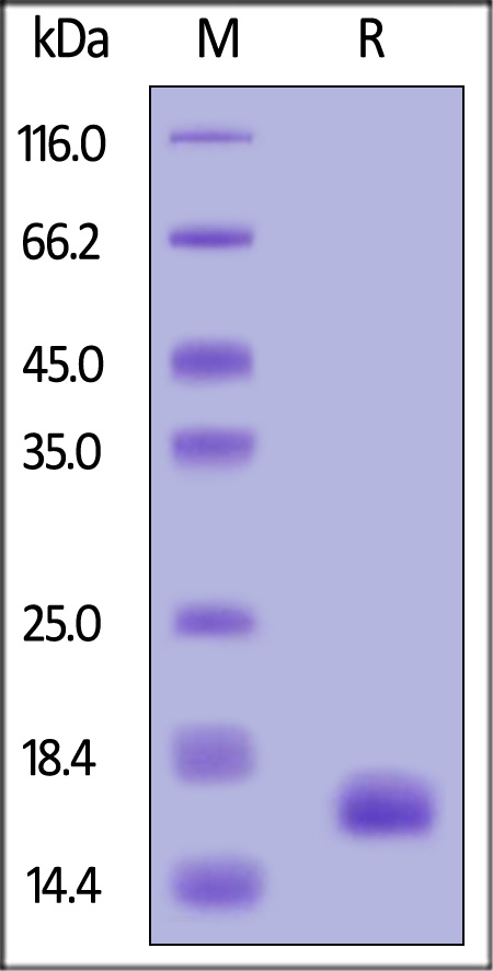
Human IL-2 Protein, Tag Free on SDS-PAGE under reducing (R) condition. The gel was stained with Coomassie Blue. The purity of the protein is greater than 95% (With Star Ribbon Pre-stained Protein Marker).

SEC-MALS

IL-2 SEC-MALS

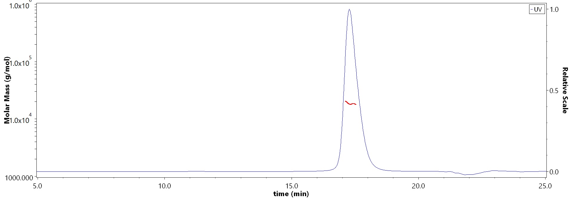
The purity of Human IL-2 Protein, Tag Free (Cat. No. IL2-H5215) is more than 95% and the molecular weight of this protein is around 14-22 kDa verified by SEC-MALS.

Report


活性(Bioactivity)-Bioactivity CELL BASE

IL-2 CELL

Human IL-2 Protein, Tag Free (Cat. No. IL2-H5215) stimulates proliferation of CTLL-2 cells. The specific activity of Human IL-2 Protein, Tag Free is > 0.60 x 10^7 IU/mg, which is calibrated against human IL-2 WHO International Standard (NIBSC code: 86/500) (QC tested).

Protocol


活性(Bioactivity)-Cell Proliferation Assay

IL-2 CELL PROLIFERATION

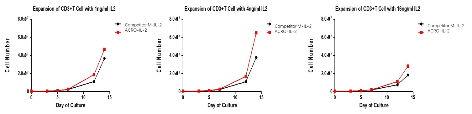
Human IL-2 Protein, Tag Free (Cat. No. IL2-H5215) has higher bioactivity than imported competitors when activates T cell proliferation with CD3/CD28 Activation Magnetic Beads.

 
评论(7)
  1. 176XXXXXXX7
  2. 6人赞
  3. ACRO的IL-2系列的蛋白用了好几种tagd~每种标签都很奈斯,结合活性结果重现性很好,官方的PROTOCOL条件很精准~基本重现没有太多误差。好评
  4. 2023-2-22
  1. 159XXXXXXX4
  2. 3人赞
  3. 今天使用了商品,实验效果不错哦,结果和预期的比较符合,后续会针对结果继续分析讨论,看看是否再次进行重复实验。
  4. >
  5. 2023-3-13
  1. 153XXXXXXX3
  2. 2人赞
  3. 我们用此蛋白用于小鼠体内动物实验,探讨重组IL2在小鼠体内实验的最大耐受剂量以及有效性,作为一个对标分子,数据持续更新中,并且结果满足期待,Acro蛋白质量很高,值得信赖,推荐。
  4. 2023-7-6
 
ACRO质量管理体系


背景(Background)

Interleukin-2 (IL-2) is an interleukin, a type of cytokine immune system signaling molecule, which is a leukocytotrophic hormone that is instrumental in the body's natural response to microbial infection and in discriminating between foreign (non-self) and self. IL-2 mediates its effects by binding to IL-2 receptors, which are expressed by lymphocytes, the cells that are responsible for immunity. Mature human IL-2 shares 56% and 66% aa sequence identity with mouse and rat IL-2, respectively. Human and mouse IL-2 exhibit crossspecies activity. The receptor for IL-2 consists of three subunits that are present on the cell surface in varying preformed complexes. IL-2 is also necessary during T cell development in the thymus for the maturation of a unique subset of T cells that are termed regulatory T cells (T-regs). After exiting from the thymus, T-Regs function to prevent other T cells from recognizing and reacting against "self antigens", which could result in "autoimmunity". T-Regs do so by preventing the responding cells from producing IL-2. Thus, IL-2 is required to discriminate between self and non-self, another one of the unique characteristics of the immune system.

文献引用(Citations)


前沿进展

Effect and Mechanism of Vitamin D on Inflammatory Factors and Neutrophil Activity in Preterm Placenta of Rats Induced by LPS
Yang, Chen, Lv
Cell Biochem Biophys (2025)
Abstract: To investigate the impact mechanisms of vitamin D on inflammatory factors and neutrophil activity in preterm pregnant rats. 24 pregnant rats were selected as the research objects and randomly divided into control group, LPS group and LPS + VD group, with 8 rats in each group. On the second day of pregnancy, the LPS + VD group was injected intraperitoneally with 50 mg/L vitamin D30.2 mL, and the LPS group and the control group were injected with the same amount of 0.9% NaCl twice a day. On the seventh day of pregnancy, the LPS group and the LPS + VD group were injected with 0.2 mL LPS into the tail vein to establish a preterm labor model induced by infection. The control group was injected with the same amount of physiological saline into the tail vein. Placental tissues from rats in the LPS + VD group and the LPS group were collected, and the expression levels of inflammatory factors TGF-β1, TNF-α, and VDBP were detected by immunohistochemistry. At the same time, serum IL-2 concentration was measured by ELISA and radioimmunoassay, the activity of neutrophils was evaluated by flow cytometry, and the expression of Hippo-YAP signaling pathway protein was detected by Western blot. Compared with the control group, the content of TNF-α, VDBP, and TGF-β1 in placenta in LPS group were higher than that in the control group (P < 5); Compared with the LPS group, the contents of TNF-α, BP and TGF-β1 in the LPS+VD group were significantly reduced,(P < 0.05); Compared with the control group, the serum IL-2 concentration in the LPS group was significantly higher than that in the control group (P < 0.05); Compared with the LPS group, the serum IL-2 concentration in the LPS + VD group decreased significantly (P < 0.05); Compared with the control group, the neutrophil ratio and absolute neutrophil value in the LPS group were higher than those in the control group (P < 0.05); Compared with the LPS group, the neutrophil ratio and absolute value of neutrophils decreased (P < 0.05); compared with the control group, the expression levels of YAP and P-YAP protein in the LPS group increased (P < 0.05); compared with the LPS group, the expression levels of YAP and P-YAP protein in the LPS + VD group decreased (P < 0.05). Vitamin D can improve the immune status of preterm pregnant mice by inhibiting the expression of placental inflammatory factors.© 2025. The Author(s), under exclusive licence to Springer Science+Business Media, LLC, part of Springer Nature.
Serum Interleukin Levels Predict Occurrence of Acute Radiation Pneumonitis and Overall Survival in Thoracic Tumours
Zhang, Shen, Li et al
Clin Invest Med (2025) 48 (1), 29-38
Abstract: Radiation-induced lung injury (RILI) is a significant adverse effect of thoracic radiotherapy, potentially impacting patient prognosis. The risk factors for acute radiation pneumonitis (RP) have not been fully clarified. The present study evaluated the predictive value of serum interleukins (ILs) in the occurrence of RP and overall survival in patients with thoracic cancers.This single-centre retrospective observational study enrolled 435 thoracic cancer patients who underwent chest radiation therapy. Serum levels of IL-1β, IL-2, IL-4, IL-5, IL-6, IL-8, IL-10, IL-12p70, IL-17, TNF-α, IFN-γ, IFN-α were measured by cytometric bead array before radiotherapy. The relationship between clinical characteristics, serum IL levels and the occurrence of RP were analyzed. Cox regression and Kaplan-Meier methods were also performed to investigate the prognostic role of serum IL levels in these patients.The incidence of RP in these patients was 17.01%. Elevated serum levels of IL-1β, IL-2, IL-4, IL-6, IL-8, IL-10, IL-12p70, TNF-α, IFN-α were all associated with the occurrence of RP. High levels of IL-1β, IL-4, and IL-12p70 were correlated with more severe pneumonitis. Univariate and multivariate logistic regression analysis identified serum IL-6 level as an independent prognostic factor in patients receiving thoracic radiotherapy.Serum interleukin levels are linked to the development of acute RP in patients receiving thoracic radiotherapy. Serum IL-6 could serve as a valuable biomarker in identifying patients at high risk for RP, potentially guiding individualized therapeutic strategies and improving patient management in radiotherapy. Future research should focus on validating IL-6's role in larger cohorts and exploring its integration into clinical practice for the early prediction of RILI.
Upregulated TCF1+ Treg Cells With Stronger Function in Systemic Lupus Erythematosus Through Activation of the Wnt-β-Catenin
Zeng, Gao, Bahabayi et al
Immunology (2025)
Abstract: The role of T-cell factor 1 (TCF1) in human regulatory T cells (Treg) and its clinical significance in systemic lupus erythematosus (SLE) remain unclear. Through bioinformatics analysis and flow cytometry, the Tcf7 gene and TCF1 protein were found to be highly expressed in Treg cells. TCF1+ Treg cells exhibited increased expression of CTLA4 and LAG3 and higher IL-10 secretion than TCF1- Treg cells. Circulating TCF1+ Treg cells were elevated and displayed increased inhibitory markers in SLE patients. The Wnt-β-catenin pathway was activated in TCF1+ Treg cells in SLE patients. The addition of XAV939 impaired the function of TCF1+ Treg cells. Clinically, TCF1+ Treg cells were not only related to CRP, ESR and IL-2, but also could differentiate SLE patients from healthy controls, primary Sjögren's syndrome patients and rheumatoid arthritis patients. In conclusion, the increased TCF1+ Treg cells in SLE patients indicate a stronger suppressive function for the activated Wnt-β-catenin pathway and help screening and assisting in the diagnosis of SLE patients.© 2025 John Wiley & Sons Ltd.
Analysis of the therapeutic effect of acitretin capsules combined with cooling blood and detoxifying formula on psoriasis hemorrhagic fever and its impact on the gut microbiota of patients
Zhou, Li, Liu et al
Medicine (Baltimore) (2025) 104 (12), e41502
Abstract: This study evaluates the therapeutic effect of acitretin capsules combined with a cooling blood and detoxifying formula (CBDF) on psoriasis hemorrhagic fever and its impact on the gut microbiota (GM). Seventy patients diagnosed with psoriasis hemorrhagic fever between January 2022 and April 2023 were divided into 2 groups: an experimental group (EG) treated with acitretin capsules and the CBDF, and a control group treated with acitretin capsules alone. The study assessed immune inflammatory responses, symptom improvement, traditional Chinese medicine symptom scores, and changes in GM diversity and abundance before and after treatment. After 2 months, the EG showed significant reductions in interleukin-2 receptor levels (from 813.2 ± 201.5-402.3 ± 111.2 mg/L) and interleukin-6 (from 8.4 ± 3.1-3.8 ± 1.7 pg/mL). The quality of life index improved by 6.12 points in the EG, which was significantly better than the control group (P < .05). PASI scores decreased in both groups, with the EG showing greater improvement (PASI before treatment: 23.1 ± 4.3; after 1 month: 15.6 ± 3.8; after 2 months: 10.4 ± 3.3). The EG also showed a significant increase in GM diversity after treatment (P < .05). Acitretin capsules combined with the CBDF is effective in treating psoriasis hemorrhagic fever and positively affects the GM, offering a potential new treatment approach.Copyright © 2025 the Author(s). Published by Wolters Kluwer Health, Inc.
Showing 1-4 of 91326 papers.
Powered by BizGenius
 
 
货号/价格
文档
联系电话:
+86 400-682-2521(全国)
010-53681107(北京)
021-50850665(上海)
运输方式
订单邮箱:
order.cn@acrobiosystems.com
技术支持邮箱:
tech.cn@acrobiosystems.com
IL-2靶点信息
英文全称:Interleukin-2
中文全称:白细胞介素-2
种类:Homo sapiens
上市药物数量:1详情
临床药物数量:39详情
最高研发阶段:批准上市
查看更多信息
前沿进展
点击查看详细

消息提示

请输入您的联系方式,再点击提交!

确定