登录 | 注册    关注公众号  
微信公众号
搜索
 >  Protein>VP1 >VP1-A5143

AAV2 VP1, Recombinant Protein

分子别名(Synonym)

capsid protein

表达区间及表达系统(Source)

AAV2 VP1, Recombinant Protein (VP1-A5143) is expressed from E. coli cells. It contains AA Met1 - Leu735 (Accession # P03135-1).

Predicted N-terminus: Met

Request for sequence

蛋白结构(Molecular Characterization)

VP1 Structure

This protein carries a polyhistidine tag at the N-terminus.

The protein has a calculated MW of 84.0 kDa. The protein migrates as 45-90 kDa when calibrated against Star Ribbon Pre-stained Protein Marker under reducing (R) condition (SDS-PAGE).

内毒素(Endotoxin)

Less than 1.0 EU per μg by the LAL method.

纯度(Purity)

>85% as determined by SDS-PAGE.

制剂(Formulation)

Supplied as 0.2 μm filtered solution in 20 mM Tris, 0.5 M NaCl, 0.5 M Arginine, pH8.0 with trehalose as protectant.

Contact us for customized product form or formulation.

运输(Shipping)

This product is supplied and shipped with dry ice, please inquire the shipping cost.

存储(Storage)

Please avoid repeated freeze-thaw cycles.

This product is stable after storage at:

  1. The product MUST be stored at -70°C or lower upon receipt;
  2. -70°C for 3 months under sterile conditions.

质量管理控制体系(QMS)

  1. 质量管理体系(ISO, GMP)
  2. 质量优势
  3. 质控流程
 

电泳(SDS-PAGE)

VP1 SDS-PAGE

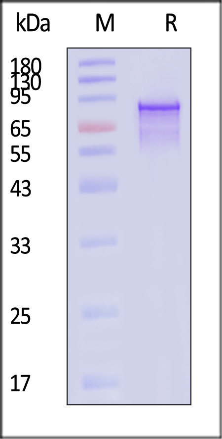
AAV2 VP1, Recombinant Protein on SDS-PAGE under reducing (R) condition. The gel was stained with Coomassie Blue. The purity of the protein is greater than 85% (With Star Ribbon Pre-stained Protein Marker).


活性(Bioactivity)-ELISA

VP1 ELISA

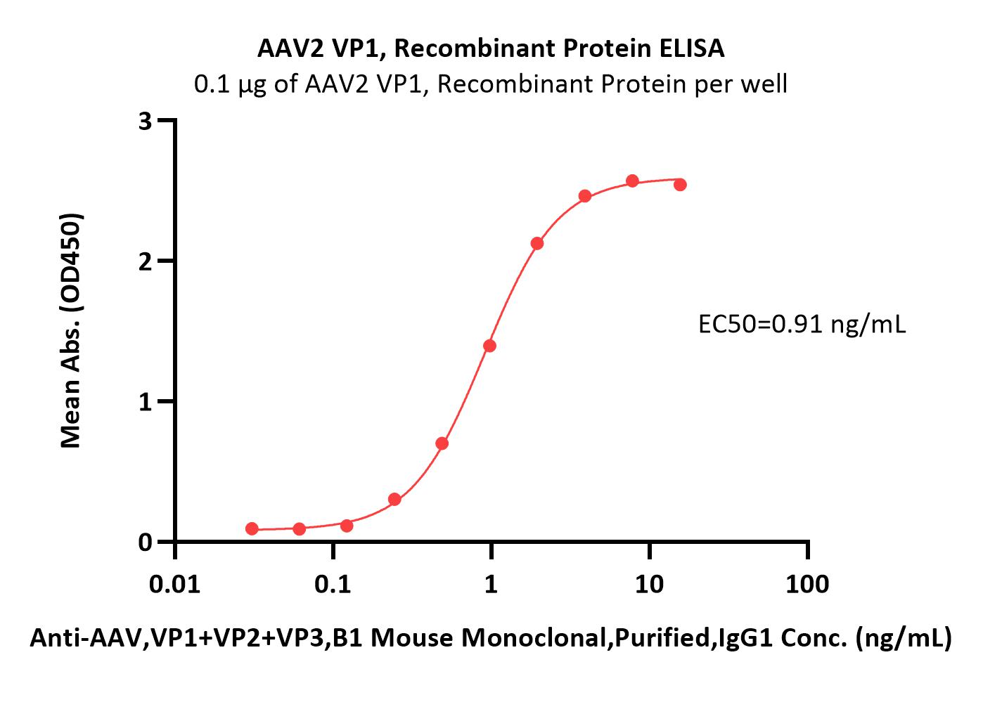
Immobilized AAV2 VP1, Recombinant Protein (Cat. No. VP1-A5143) at 1 μg/mL (100 μL/well) can bind Anti-AAV,VP1+VP2+VP3,B1 Mouse Monoclonal,Purified,IgG1 with a linear range of 1-31 ng/mL (QC tested).

Protocol

 
评论(0)
 
ACRO质量管理体系


背景(Background)

Adeno-associated virus is a single-stranded DNA virus and the current scientific consensus is that it does not cause any human disease. It consists of a protein capsid (CAPside) and a 4.7 KB length single stranded DNA genome. The protein capsid consists of three subunits, VP1, VP2, and VP3.


前沿进展

A single amino acid variant in the variable region I of AAV capsid confers liver detargeting
Xing, Xu, Reil et al
bioRxiv (2025)
Abstract: AAV capsid serotypes isolated from nature have been widely used in gene delivery and gene therapy. Recently, more than 1,000 distinct AAV capsids were identified from human clinical samples by high-throughput, long-read DNA sequencing (Hsu HL et al. Nature Communications 2020). In this study, we tap into this broad natural biodiversity of AAV capsids to develop liver-tropic AAV capsids. We initially screened a subset of variants derived from AAV8 (n=159) for packaging efficiency. The top 30% of these variants were subjected to a barcoded vector library screen in mice and ferrets for their ability to mediate liver gene transfer. Although no variant surpassed AAV8 for liver targeting, several exhibited a liver detargeting phenotype. Among these, we focused on the N271D variant (AAV8 VP1 numbering), located in the variable region I (VR-1), which has been previously implicated in influencing liver tropism (Cabanes-Creus M et al. Molecular Therapy Methods & Clinical Development 2021; Zinn E et al. Cell Reports Medicine 2022). The liver detargeting phenotype of AAV8.N271D was confirmed by single vector administration in mice. Additionally, we grafted the N271D variant onto AAV9 and MyoAAV capsids (N270D by AAV9 VP1 numbering). The AAV9.N270D and MyoAAV.N270D vectors showed a similar liver-detargeting phenotype, although muscle targeting was moderately reduced. This study reinforces the important role of VR-1 in modulating liver tropism, and highlights the potential of engineering the VR-1 residues to mitigate liver gene transfer and associated toxicity.
Enterovirus VP1 protein and HLA class I hyperexpression in pancreatic islet cells of organ donors with type 1 diabetes
Rodriguez-Calvo, Laiho, Oikarinen et al
Diabetologia (2025)
Abstract: Earlier studies of pancreases from donors with type 1 diabetes demonstrated enteroviral capsid protein VP1 in beta cells. In the context of a multidisciplinary approach undertaken by the nPOD-Virus group, we assessed VP1 positivity in pancreas and other tissues (spleen, duodenum and pancreatic lymph nodes) from 188 organ donors, including donors with type 1 diabetes and donors expressing autoantibody risk markers. We also investigated whether VP1 positivity is linked to the hyperexpression of HLA class I (HLA-I) molecules in islet cells.Organ donor tissues were collected by the Network for Pancreatic Organ Donors with Diabetes (nPOD) from donors without diabetes (ND, n=76), donors expressing a single or multiple diabetes-associated autoantibodies (AAb+, n=20; AAb++, n=9) and donors with type 1 diabetes with residual insulin-containing islets (T1D-ICIs, n=41) or only insulin-deficient islets (T1D-IDIs, n=42). VP1 was assessed using immunohistochemistry (IHC) and HLA-I using IHC and immunofluorescence, in two independent laboratories. We determined assay concordance across laboratories and overall occurrence of positive assays, on a case-by-case basis and between donor groups.Islet cell VP1 positivity was detected in most T1D-ICI donors (77.5%) vs only 38.2% of ND donors (p<0.001). VP1 positivity was associated with HLA-I hyperexpression. Of those donors assessed for HLA-I and VP1, 73.7% had both VP1 immunopositivity and HLA-I hyperexpression (p<0.001 vs ND). Moreover, VP1+ cells were detected at higher frequency in donors with HLA-I hyperexpression (p<0.001 vs normal HLA-I). Among VP1+ donors, the proportion with HLA-I hyperexpression was significantly higher in the AAb++ and T1D-ICI groups (94.9%, p<0.001 vs ND); this was not restricted to individuals with recent-onset diabetes. Critically, for all donor groups combined, HLA-I hyperexpression occurred more frequently in VP1+ compared with VP1- donors (45.8% vs 16%, p<0.001).We report the most extensive analysis to date of VP1 and HLA-I in pancreases from donors with preclinical and diagnosed type 1 diabetes. We find an association of VP1 with residual beta cells after diagnosis and demonstrate VP1 positivity during the autoantibody-positive preclinical stage. For the first time, we show that VP1 positivity and HLA-I hyperexpression in islet cells are both present during the preclinical stage. While the study of tissues does not allow us to demonstrate causality, our data support the hypothesis that enterovirus infections may occur throughout the natural history of type 1 diabetes and may be one of multiple mechanisms driving islet cell HLA-I hyperexpression.© 2025. The Author(s).
Joint analysis of the nPOD-Virus Group data: the association of enterovirus with type 1 diabetes is supported by multiple markers of infection in pancreas tissue
Richardson, Rodriguez-Calvo, Laiho et al
Diabetologia (2025)
Abstract: Previous pathology studies have associated enterovirus infections with type 1 diabetes by examining the enterovirus capsid protein 1 (VP1) in autopsy pancreases obtained near diabetes diagnosis. The Network for Pancreatic Organ Donors with Diabetes (nPOD) has since obtained pancreases from organ donors with type 1 diabetes (with broad age and disease duration) and donors with disease-associated autoantibodies (AAbs), the latter representing preclinical disease. Two accompanying manuscripts from the nPOD-Virus Group report primary data from a coordinated analysis of multiple enterovirus indices. We aimed to comprehensively assess the association of multiple enterovirus markers with type 1 diabetes.The nPOD-Virus Group examined pancreases from 197 donors, recovered between 2007 and 2019, classified into five groups: donors with type 1 diabetes, with residual insulin-containing islets (T1D-ICI group, n=41) or with only insulin-deficient islets (T1D-IDI, n=42); donors without diabetes who are AAb-negative (ND, n=83); and rare donors without diabetes expressing a single AAb (AAb+, n=22) or multiple AAbs (AAb++, n=9). We assessed the overall association of multiple indicators of enterovirus infection, case-by-case and between donor groups, as well as assay agreement and reproducibility, using various statistical methods. We examined data from 645 assays performed across 197 nPOD donors.Detection of enterovirus indices by independent laboratories had high reproducibility, using both enterovirus-targeted and unbiased methods. T1D-ICI donors had significantly higher (p<0.001) proportions of positive assay outcomes (58.4%) vs T1D-IDI (10.3%), ND (17.8%) and AAb-positive donors (AAb+ 24.6%; AAb++ 35.0%). Head-to-head comparisons revealed increased proportions of donors positive in two independent assays among T1D-ICI vs ND donors (VP1/HLA class I [HLA-I], p<0.0001; VP1/enterovirus-specific RT-PCR (EV-PCR), p=0.076; EV-PCR/HLA-I, p=0.016; proteomics/HLA-I, p<0.0001; VP1/proteomics, p=0.06). Among 110 donors examined for three markers (VP1, EV-PCR and HLA-I), 83.3% of T1D-ICI donors were positive in two or more assays vs 0% of ND (p<0.001), 26.7% of AAb+ (p=0.006), 28.6% of AAb++ (p=0.023) and 0% of T1D-IDI (p<0.001) donors.The nPOD-Virus Group conducted, to date, the largest and most comprehensive analysis of multiple indices of pancreatic enterovirus infections in type 1 diabetes; these were more prevalent in T1D-ICI and AAb++ donors than in other groups. Their preferential detection of these indices in donors with residual beta cells and autoimmunity implicates enterovirus infections across disease progression stages and supports a contribution to beta cell loss, directly or indirectly, even after diagnosis. The relatively small number of infected cells and the low amount of viral RNA support the existence of non-acute, low level, possibly persistent enterovirus infections in the pancreas.© 2025. The Author(s).
Molecular identification of novel duck associated Chapparvovirus in ducks, first report from China
Man, Li, Xu et al
Poult Sci (2025) 104 (5), 104984
Abstract: Duck Associated Chapparvovirus (DaChPV) is a newly discovered virus within the Chaphamparvovirus genus, and first identified from Canadian wild ducks in 2021. In this study, DaChPV DNA was detected in 14 out of 137 tissues samples collected from diarrhea ducks across various provinces in China. Subsequently, eight complete genome sequences were amplified using overlapping primers and sequenced. Comparative analysis revealed that the amino acid (aa) sequences of NS1 and VP1 from these eight DaChPV strains shared identity with reference strains, ranging from 81.43 % to 98.81 % for NS1 and 76.95 % to 98.39 % for VP1, respectively. Phylogenetic analysis of the genome sequences showed that the newly identified DaChPVs and reference DaChPV strains formed an independent cluster, indicating a close genetic relationship. The strains of AH2301, HN2201 and HN2301 identified in this study were classified as belonging to Duck Associated Chaphamapavovirus 1. In contrast, AH2401, HN2401, and SD2301 were grouped with Duck Associated Chaphamapavovirus 2. The remaining two strains, HeB2201 and HeB2401, may represent variant strains that cluster independently, which is further supported by the evolutionary tree results of VP1 and NS1. Additionally, inter-type recombinations were predicted for these DaChPV strains. The contained multiple specific mutation sites, including 63, 76, 78, 303, and 305 that located on the predicted antigenic epitopes. This research firstly determined the evolutionary trends of DaChPV in China, offering valuable insights for understanding of spread, evolution, and molecular epidemiology of DaChPV on a global scale.Copyright © 2025. Published by Elsevier Inc.
Showing 1-4 of 8164 papers.
Powered by BizGenius
 
 
货号/价格
文档
联系电话:
+86 400-682-2521(全国)
010-53681107(北京)
021-50850665(上海)
运输方式
订单邮箱:
order.cn@acrobiosystems.com
技术支持邮箱:
tech.cn@acrobiosystems.com
VP1靶点信息
英文全称:Capsid protein
中文全称:衣壳蛋白
种类:
上市药物数量:1详情
临床药物数量:27详情
最高研发阶段:批准上市
查看更多信息
前沿进展
点击查看详细
相关产品
View All VP1

消息提示

请输入您的联系方式,再点击提交!

确定