Integrated Analysis of Gene Expression and Immune Cell Infiltration Reveals Dysregulated Genes and miRNAs in Acute Kidney InjuryZhang, Gong, Lu
et alMol Biotechnol (2024)
Abstract: Acute Kidney Injury (AKI) is a multifaceted condition characterised by rapid deterioration of renal function, often precipitated by diverse etiologies. A comprehensive understanding of the molecular underpinnings of AKI is pivotal for identifying potential diagnostic markers and therapeutic targets. This study utilised bioinformatics to elucidate gene expression and immune infiltration in AKI. Publicly available mRNA and miRNA datasets were harnessed to discern differentially expressed genes (DEGs) and miRNAs in AKI. The CIBERSORT algorithm was employed to quantify immune cell infiltration in AKI samples. Functional enrichment analyses were conducted to unravel the implicated biological processes. Furthermore, the expression of identified genes and miRNAs was validated by quantitative real-time PCR in an AKI model. Our study revealed significant dysregulation of three genes (Aspn, Clec2h, Tmigd1) and two miRNAs (mmu-miR-21a-3p, mmu-miR-223-3p) in AKI, each with p < 0.0001. These molecular markers are implicated in immune responses, tissue remodelling, and inflammation. We observed notable disturbances in specific immune cells, including activated and immature dendritic cells, M1 macrophages, and subsets of T cells (Treg, Th1, Th17). These alterations correlated significantly with AKI pathology, with dendritic cells and M1 macrophages showing p < 0.01, and T cell subsets demonstrating p < 0.05. These results highlight the intricate involvement of the immune system in AKI and indicate significant enrichment of pathways related to immune response, inflammation, and tissue remodelling, pointing to their pivotal roles in AKI pathophysiology. Our study underscored the significance of immune cell infiltration and dysregulated gene and miRNA expression in AKI. The identified genes (Clec2h, Aspn, and Tmigd1) and miRNAs (mmu-miR-21a-3p and mmu-miR-223-3p) offer potential diagnostic markers and therapeutic avenues for AKI. Subsequent investigations targeting these genes and miRNAs, along with the elucidated pathways, may augment the clinical management and outcomes for AKI patients.© 2024. The Author(s), under exclusive licence to Springer Science+Business Media, LLC, part of Springer Nature.
Integrative analysis of the roles of lncRNAs and mRNAs in ischaemic preconditioning to alleviate liver ischaemia-reperfusion injury in miceHua, Xu, Li
et alBiochem Biophys Res Commun (2022) 627, 30-38
Abstract: The objective of our study was to elucidate the possible underlying mechanism for the protective effect of ischaemic preconditioning (IPC) against ischaemia-reperfusion (I/R) injury and to provide new research perspectives of long non-coding RNAs (lncRNAs). In this study, serum and liver tissue samples were collected to measure indexes of liver injury from a mouse liver model in sham, I/R injury and I/R + IPC groups. Furthermore, liver samples from 5 randomly selected mice per group were extracted and subjected to the microarray and subsequent bioinformatics analysis. IPC ameliorated liver damage by lowered liver transaminase levels and pro-inflammatory cytokines. A total of 167 lncRNAs and 108 messenger RNAs (mRNAs) were significantly differentially expressed genes (DEGs) between the I/R + IPC and I/R groups. Gene Ontology (GO) analysis revealed that these genes were mainly related to unfolded proteins, responses to topologically incorrect proteins, responses to temperature stimuli, protein folding and protein refolding. Kyoto Encyclopaedia of Genes and Genomes (KEGG) pathway analysis indicated that the DEGs were enriched in the following pathways: protein processing in the endoplasmic reticulum; antigen processing and presentation; and fructose and mannose metabolism. Additionally, the 7 selected DEGs (Hspa1ab, Chka, Clec2h, Mvd, Adra1a, AK085737 and AK088966) were validated in modules of the lncRNA-mRNA weighted coexpression network, which agreed with the qRT-PCR and chip data. And the identified differentially expressed lncRNAs and mRNAs may provide new clues to understand the pivotal pathophysiological mechanism by which IPC alleviates I/R-caused liver damage.Copyright © 2022 Elsevier Inc. All rights reserved.
Clr-f expression regulates kidney immune and metabolic homeostasisZein, Abou-Samra, Scur
et alSci Rep (2022) 12 (1), 4834
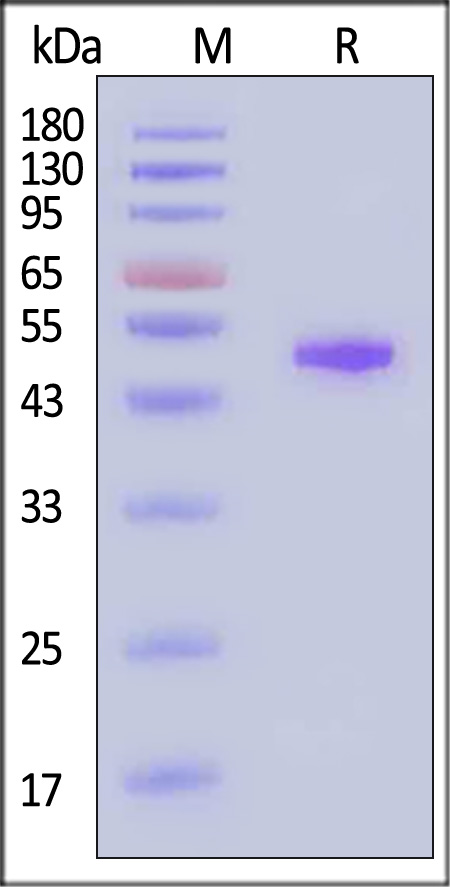
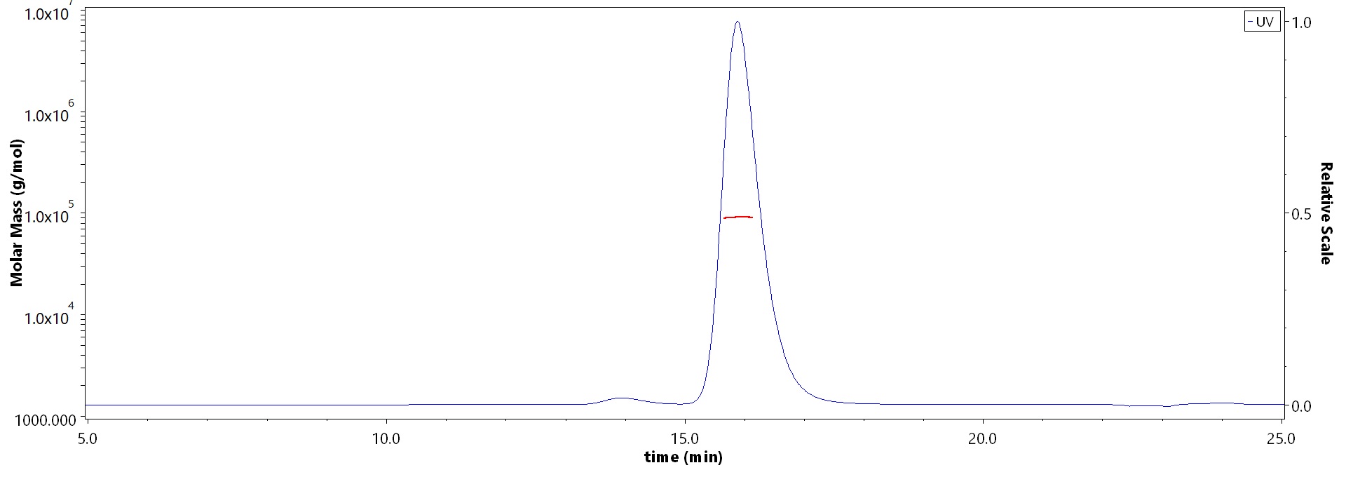
Abstract: The C-type lectin-related protein, Clr-f, encoded by Clec2h in the mouse NK gene complex (NKC), is a member of a family of immune regulatory lectins that guide immune responses at distinct tissues of the body. Clr-f is highly expressed in the kidney; however, its activity in this organ is unknown. To assess the requirement for Clr-f in kidney health and function, we generated a Clr-f-deficient mouse (Clr-f-/-) by targeted deletions in the Clec2h gene. Mice lacking Clr-f exhibited glomerular and tubular lesions, immunoglobulin and C3 complement protein renal deposits, and significant abdominal and ectopic lipid accumulation. Whole kidney transcriptional profile analysis of Clr-f-/- mice at 7, 13, and 24 weeks of age revealed a dynamic dysregulation in lipid metabolic processes, stress responses, and inflammatory mediators. Examination of the immune contribution to the pathologies of Clr-f-/- mouse kidneys identified elevated IL-12 and IFNγ in cells of the tubulointerstitium, and an infiltrating population of neutrophils and T and B lymphocytes. The presence of these insults in a Rag1-/-Clr-f-/- background reveals that Clr-f-/- mice are susceptible to a T and B lymphocyte-independent renal pathogenesis. Our data reveal a role for Clr-f in the maintenance of kidney immune and metabolic homeostasis.© 2022. The Author(s).
The Effect of Lactobacillus casei 32G on the Mouse Cecum Microbiota and Innate Immune Response Is Dose and Time DependentAktas, De Wolfe, Tandee
et alPLoS One (2015) 10 (12), e0145784
Abstract: Lactobacilli have been associated with a variety of immunomodulatory effects and some of these effects have been related to changes in gastrointestinal microbiota. However, the relationship between probiotic dose, time since probiotic consumption, changes in the microbiota, and immune system requires further investigation. The objective of this study was to determine if the effect of Lactobacillus casei 32G on the murine gastrointestinal microbiota and immune function are dose and time dependent. Mice were fed L. casei 32G at doses of 106, 107, or 108 CFU/day/mouse for seven days and were sacrificed 0.5h, 3.5h, 12h, or 24h after the last administration. The ileum tissue and the cecal content were collected for immune profiling by qPCR and microbiota analysis, respectively. The time required for L. casei 32G to reach the cecum was monitored by qPCR and the 32G bolus reaches the cecum 3.5h after the last administration. L. casei 32G altered the cecal microbiota with the predominance of Lachnospiraceae IS, and Oscillospira decreasing significantly (p < 0.05) in the mice receiving 108 CFU/mouse 32G relative to the control mice, while a significant (p < 0.05) increase was observed in the prevalence of lactobacilli. The lactobacilli that increased were determined to be a commensal lactobacilli. Interestingly, no significant difference in the overall microbiota composition, regardless of 32G doses, was observed at the 12h time point. A likely explanation for this observation is the level of feed derived-nutrients resulting from the 12h light/dark cycle. 32G results in consistent increases in Clec2h expression and reductions in TLR-2, alpha-defensins, and lysozyme. Changes in expression of these components of the innate immune system are one possible explanation for the observed changes in the cecal microbiota. Additionally, 32G administration was observed to alter the expression of cytokines (IL-10rb and TNF-α) in a manner consistent with an anti-inflammatory response.
Showing 1-4 of 4 papers.


 膜杰作
膜杰作 Star Staining