登录 | 注册    关注公众号  
微信公众号
搜索
 >  Protein>H-2Db & B2M >H2M-M82E9

Biotinylated Mouse H-2Db&B2M Monomer Protein (Peptide free, MALS verified)

分子别名(Synonym)

H-2Db & B2M

表达区间及表达系统(Source)

Biotinylated Mouse H-2Db&B2M Monomer Protein (H2M-M82E9) is expressed from human 293 cells (HEK293). It contains AA Gly 25 - Val 309 (H-2Db) & Ile 21 - Met 119 (B2M) (Accession # P01899 (H-2Db) & P01887 (B2M)).

Predicted N-terminus: Gly 25 & Ile 21

Request for sequence

蛋白结构(Molecular Characterization)

This protein carries a polyhistidine tag at the C-terminus, followed by an Avi tag (Avitag™).

The protein has a calculated MW of 40.1 kDa and 15.7 kDa. The protein migrates as 50-55 kDa and 16 kDa when calibrated against Star Ribbon Pre-stained Protein Marker under reducing (R) condition (SDS-PAGE) due to glycosylation.

标记(Labeling)

Biotinylation of this product is performed using Avitag™ technology. Briefly, the single lysine residue in the Avitag is enzymatically labeled with biotin.

蛋白标记度(Protein Ratio)

Passed as determined by the HABA assay / binding ELISA.

内毒素(Endotoxin)

Less than 1.0 EU per μg by the LAL method.

纯度(Purity)

>95% as determined by SDS-PAGE.

>90% as determined by SEC-MALS.

制剂(Formulation)

Lyophilized from 0.22 μm filtered solution in PBS, pH7.4 with trehalose as protectant.

Contact us for customized product form or formulation.

重构方法(Reconstitution)

Please see Certificate of Analysis for specific instructions.

For best performance, we strongly recommend you to follow the reconstitution protocol provided in the CoA.

存储(Storage)

For long term storage, the product should be stored at lyophilized state at -20°C or lower.

Please avoid repeated freeze-thaw cycles.

This product is stable after storage at:

  1. -20°C to -70°C for 12 months in lyophilized state;
  2. -70°C for 3 months under sterile conditions after reconstitution.

质量管理控制体系(QMS)

  1. 质量管理体系(ISO, GMP)
  2. 质量优势
  3. 质控流程
 

电泳(SDS-PAGE)

H-2Db & B2M SDS-PAGE

Biotinylated Mouse H-2Db&B2M Monomer Protein on SDS-PAGE under reducing (R) condition. The gel was stained with Coomassie Blue. The purity of the protein is greater than 95% (With Star Ribbon Pre-stained Protein Marker).

SEC-MALS

H-2Db & B2M SEC-MALS

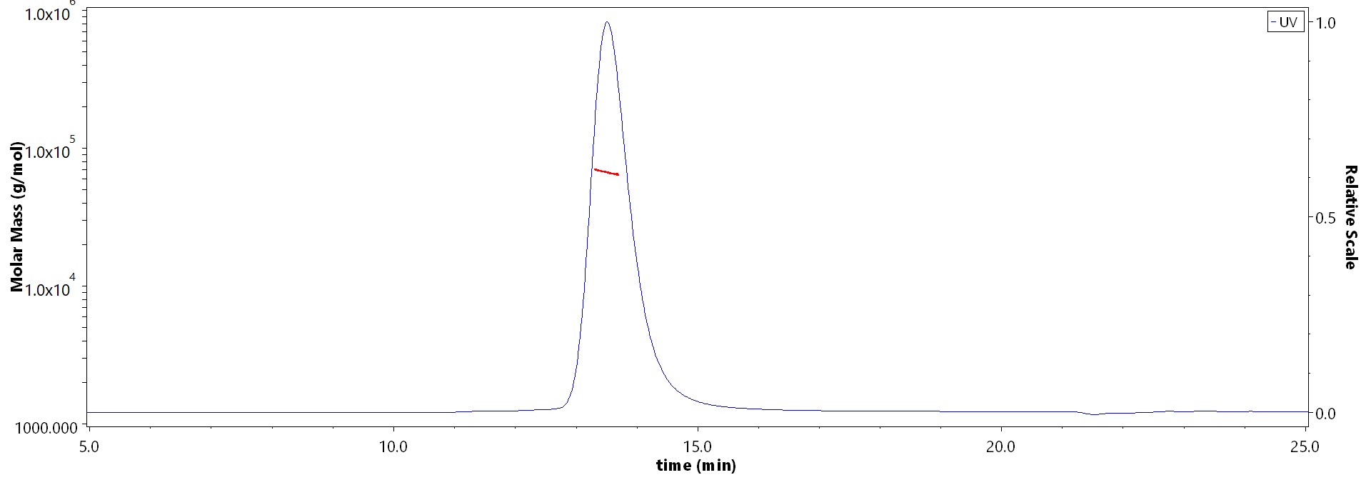
The purity of Biotinylated Mouse H-2Db&B2M Monomer Protein (Cat. No. H2M-M82E9) is more than 90% and the molecular weight of this protein is around 55-70 kDa verified by SEC-MALS.

Report


活性(Bioactivity)-ELISA

H-2Db & B2M ELISA

Immobilized Biotinylated Mouse H-2Db&B2M Monomer Protein (Cat. No. H2M-M82E9) at 1 μg/mL (100 μL/well) on streptavidin (Cat. No. STN-N5116) precoated (0.5 μg/well) plate can bind Mouse H-2Kb/H-2Db Monoclonal Antibody with a linear range of 0.001-0.156 μg/mL (QC tested).

Protocol

H-2Db & B2M ELISA

Immobilized Biotinylated Mouse H-2Db&B2M Monomer Protein (Cat. No. H2M-M82E9) at 1 μg/mL (100 μL/well) on streptavidin (Cat. No. STN-N5116) precoated (0.5 μg/well) plate can bind Anti-B2M Antibody, Human IgG1 with a linear range of 0.5-16 ng/mL (Routinely tested).

Protocol

 
评论(0)
 
ACRO质量管理体系


前沿进展

Establishment and characterization of a mouse tumor cell line with irreversible downregulation of MHC class I molecules
Lhotakova, Grzelak, Polakova et al
Oncol Rep (2019) 42 (6), 2826-2835
Abstract: In the majority of human tumors, downregulation of major histocompatibility complex class I (MHC‑I) expression contributes to the escape from the host immune system and resistance to immunotherapy. Relevant animal models are therefore needed to enhance the efficacy of cancer immunotherapy. As loss of β‑2 microglobulin expression results in irreversible downregulation of surface MHC‑I molecules in various human tumors, the β‑2 microglobulin gene (B2m) was deactivated in a mouse oncogenic TC‑1 cell line and a TC‑1/dB2m cell line that was negative for surface MHC‑I expression was derived. Following stimulation with interferon γ, MHC‑I heavy chains, particularly the H‑2Db molecules, were found to be expressed at low levels on the cell surface, but without β‑2 microglobulin. B2m deactivation in TC‑1/dB2m cells led to reduced proliferation and tumor growth. These cells were insensitive to DNA vaccination and only weakly responsive to combined immunotherapy with a DNA vaccine and the ODN1826 adjuvant. In vivo depletion demonstrated that NK1.1+ cells were involved in both reduced tumor growth and an antitumor effect of immunotherapy. The number of immune cells infiltrating TC‑1/dB2m‑induced tumors was comparable with that in tumors developing from TC‑1/A9 cells characterized by reversible MHC‑I downregulation. However, the composition of the cell infiltrate was different and, most importantly, infiltration with immune cells was not increased in TC‑1/dB2m tumors after immunotherapy. Therefore, the TC‑1/dB2m cell line represents a clinically relevant tumor model that may be used for enhancement of cancer immunotherapy.
Cell-surface expression of H-2Db requires N-linked glycans
Fraser, Allen, Flavell et al
Immunogenetics (1987) 26 (1-2), 31-5
Abstract: The question of whether beta-2 microglobulin (B2m)-independent expression of the mouse major histocompatibility complex (MHC) antigen H-2Db results from the atypical glycosylation pattern associated with this MHC antigen (i.e., three glycans instead of two) has been addressed. Cell-surface expression of transfected H-2Db in the B2m deficient cell line R1E was completely abolished by the drug tunicamycin (Tm). Introduction of a functional B2m gene by transfection did not re-establish cell-surface expression of Db in the presence of Tm. Tm had no effect, however, on the expression of a truncated Db molecule lacking the alpha 1 and alpha 2 domains which is glycosylated at amino acid position 256, suggesting that the Db molecule, unlike other class I antigens, possesses an unstable conformation in the alpha 1 and/or alpha 2 domains which requires the attachment of glycans before it is transported to the cell surface. Once attached, however, glycans may confer a stable alpha 1/alpha 2 conformation apparently peculiar to Db which allows cell-surface expression in the absence of B2m.
Antigenic determinants shared between HLA-A, -B, -C antigens and H-2 class I molecules modified by bovine beta-2 microglobulin
Mierau, Robinson, Sanderson et al
Immunogenetics (1987) 26 (6), 351-5
Abstract: The specificity of the mouse class I-specific antibody COB6-3 was examined in detail. It was found to react with the mouse class I molecules H-2Db, Kd, and Qa-2, and with human HLA-A, -B, -C antigens. The specificity pattern of COB6-3, despite its different origin, was similar to that of the monomorphic HLA class I-specific antibody W6/32. Cross-inhibition studies show that on human cells the antigenic determinants recognized by the two antibodies are situated close together and may be identical. On mouse cells, reactivity of both antibodies was generated upon replacement of mouse beta-2 microglobulin (B2m) with its bovine counterpart, but differences in specificity were observed using human B2m.
Molecular analysis of an EL4 cell line that expresses H-2Db but not H-2Kb or beta 2-microglobulin
Potter, Zeff, Schmitt-Verhulst et al
Proc Natl Acad Sci U S A (1985) 82 (9), 2950-4
Abstract: EL4/Mar is a variant cell line that expresses H-2Db but neither H-2Kb nor beta 2-microglobulin (beta 2m). Southern and RNA blot analysis and immunoprecipitation of metabolically labeled proteins established that the B2m gene(s), beta 2m mRNA, and beta 2m protein are normal in this cell line. Somatic cell hybridization showed that the defect in this cell line was in the synthesis of H-2Kb, and RNA blot analysis with an H-2Kb specific oligonucleotide established that the H-2Kb gene(s) in this cell line was not transcribed into a stable mRNA species. The apparent absence of beta 2m on the surface of this cell line suggests that there may be some feature of the H-2Db molecule that allows it to be expressed in the absence of detectable beta 2m.
Showing 1-4 of 4 papers.
Powered by BizGenius
 
 
货号/价格
产品推荐
文档
联系电话:
+86 400-682-2521(全国)
010-53681107(北京)
021-50850665(上海)
运输方式
订单邮箱:
order.cn@acrobiosystems.com
技术支持邮箱:
tech.cn@acrobiosystems.com
前沿进展
点击查看详细

消息提示

请输入您的联系方式,再点击提交!

确定