登录 | 注册    关注公众号  
微信公众号
搜索
 >  Antibody>Hemagglutinin/HA (Influenza Virus) >HA2-M692

Monoclonal Anti-Influenza A [A/Darwin/6/2021 (H3N2)] HA Antibody, Human IgG1 (7G4) (MALS verified)

抗体来源(Source)

Monoclonal Anti-Influenza A [A/Darwin/6/2021 (H3N2)] HA Antibody, Human IgG1 (7G4) is a chimeric monoclonal antibody recombinantly expressed from HEK293, which combines the variable region of a mouse monoclonal antibody with Human constant domain.

克隆号(Clone)

7G4

亚型(Isotype)

Human IgG1 | Human Kappa

偶联(Conjugate)

Unconjugated

抗体类型(Antibody Type)

Recombinant Monoclonal

种属反应性(Reactivity)

Virus

免疫原(Immunogen)

Recombinant Influenza A [A/Darwin/6/2021 (H3N2)] HA Protein is expressed from human 293 cells.

特异性(Specificity)

Specifically recognizes Influenza A (H3N2) Viruses Hemagglutinin (HA).

应用(Application)

ApplicationRecommended Usage
ELISA0.1-5 ng/mL

纯度(Purity)

>90% as determined by SDS-PAGE.

>90% as determined by SEC-MALS.

纯化(Purification)

Protein A purified / Protein G purified

制剂(Formulation)

Lyophilized from 0.22 μm filtered solution in PBS, pH7.4 with trehalose as protectant.

Contact us for customized product form or formulation.

重构方法(Reconstitution)

Please see Certificate of Analysis for specific instructions.

For best performance, we strongly recommend you to follow the reconstitution protocol provided in the CoA.

存储(Storage)

For long term storage, the product should be stored at lyophilized state at -20°C or lower.

Please avoid repeated freeze-thaw cycles.

This product is stable after storage at:

  1. -20°C to -70°C for 12 months in lyophilized state;
  2. -70°C for 3 months under sterile conditions after reconstitution.

质量管理控制体系(QMS)

  1. 质量管理体系(ISO, GMP)
  2. 质量优势
  3. 质控流程
 

交叉验证(Cross Verification)

This product is a specific antibody against
Influenza A [A/Darwin/6/2021 (H3N2)] HA Protein, His Tag (Cat. No. HA2-V52H5).
Influenza A Virus (A/Croatia/10136RV/2023) HA (H3N2) Protein, His Tag (Cat. No. H32-V52H4).
Influenza A Virus (A/District of Columbia/27/2023) HA (H3N2) Protein, His Tag (Cat. No. H32-V52H5).
No cross-reactivity in ELISA with
Influenza A [A/Shanghai/2/2013(H7N9)] HA, Fc Tag (Cat. No. HA9-V5253).
Influenza A [A/guinea fowl/Hong Kong/WF10/99(H9N2)] HA1 Protein, His Tag (Cat. No. HA1-V52H5).
Influenza A [A/guinea fowl/Hong Kong/WF10/99(H9N2)] Hemagglutinin (HA) Protein, His Tag (Cat. No. HA2-V52H7).
Influenza A [A/Hong Kong/483/97 (H5N1)] HA, His Tag (Cat. No. HA1-V5229).
Influenza A [A/Wisconsin/588/2019 (H1N1)] HA, His Tag (Cat. No. HA1-V52H3).
Influenza A [A/Darwin/9/2021 (H3N2)] HA Protein, His Tag (Cat. No. HA2-V52H6).
Influenza A [Sydney/5/2021 (H1N1)] Hemagglutinin (HA) Protein, His Tag (MALS verified) (Cat. No. HA1-V52H4).
Influenza B [Austria/1359417/2021 (B/Victoria lineage)] Hemagglutinin (HA) Protein, His Tag (Cat. No. HAE-V52H3).
Influenza B [Phuket/3073/2013 (B/Yamagata lineage)] Hemagglutinin (HA) Protein, His Tag (Cat. No. HAE-V52H4).
Influenza A [A/Bangkok/1/1979 (H3N2)] Hemagglutinin (HA) Protein, His Tag (MALS verified) (Cat. No. HA2-V52H3).
Influenza A [A/Victoria/2570/2019] Hemagglutinin (HA) Protein, His Tag (MALS verified) (Cat. No. HA1-V52H6).
Influenza A (A/Shanghai/02/2013(H7N9)) Hemagglutinin (HA) Protein, His Tag (Cat. No. HA1-V52H6).
Influenza A [Victoria/4897/2022] Hemagglutinin (HA) Protein, His Tag (Cat. No. HA9-V52H3).
Influenza A [Victoria/4897/2022] Hemagglutinin (HA) Protein, His Tag (Cat. No. HA1-V52H8).
Influenza A (turkey/Germany-MV/R2472/2014(H5N8)) HA Protein, His Tag (Cat. No. HA8-V52H3).
Influenza A (Guangdong/18SF020(H5N6)) Hemagglutinin (HA) Protein, His Tag (Cat. No. HA6-V52H3).
Influenza A (Vietnam/1194/2004(H5N1)) Hemagglutinin (HA) Protein, His Tag (Cat. No. HA1-V52H9).
Influenza A [Wisconsin/67/2022] Hemagglutinin (HA) Protein, His Tag (Cat. No. HA1-V52H7).


电泳(SDS-PAGE)

Hemagglutinin/HA (Influenza Virus) SDS-PAGE

Monoclonal Anti-Influenza A [A/Darwin/6/2021 (H3N2)] HA Antibody, Human IgG1 (7G4) on SDS-PAGE under reducing (R) condition. The gel was stained with Coomassie Blue. The purity of the protein is greater than 90% (With Star Ribbon Pre-stained Protein Marker).

SEC-MALS

Hemagglutinin/HA (Influenza Virus) SEC-MALS

The purity of Monoclonal Anti-Influenza A [A/Darwin/6/2021 (H3N2)] HA Antibody, Human IgG1 (7G4) (Cat. No. HA2-M692) is more than 90% and the molecular weight of this protein is around 135-160 kDa verified by SEC-MALS.

Report


活性(Bioactivity)-ELISA

Hemagglutinin/HA (Influenza Virus) ELISA

Immobilized Influenza A [A/Darwin/6/2021 (H3N2)] HA Protein, His Tag (Cat. No. HA2-V52H5) at 1 μg/mL (100 μL/well) can bind Monoclonal Anti-Influenza A [A/Darwin/6/2021 (H3N2)] HA Antibody, Human IgG1 (7G4) (Cat. No. HA2-M692) with a linear range of 0.1-1 ng/mL (QC tested).

Protocol

Hemagglutinin/HA (Influenza Virus) ELISA

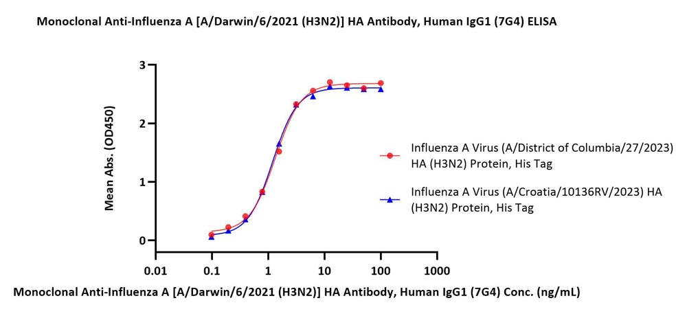
Immobilized Influenza A Virus (A/Croatia/10136RV/2023) HA (H3N2) Protein, His Tag (Cat. No. H32-V52H4), Influenza A Virus (A/District of Columbia/27/2023) HA (H3N2) Protein, His Tag (Cat. No. H32-V52H5) at 1 μg/mL (100 μL/well) can bind Monoclonal Anti-Influenza A [A/Darwin/6/2021 (H3N2)] HA Antibody, Human IgG1 (7G4) (Cat. No. HA2-M692) with a linear range of 0.1-3 ng/mL (Routinely tested).

Protocol

 
评论(0)
 
ACRO质量管理体系


背景(Background)

Neuraminidase (NA) and hemagglutinin (HA) are major membrane glycoproteins found on the surface of influenza virus. Hemagglutinin binds to the sialic acid-containing receptors on the surface of host cells during initial infection and at the end of an infectious cycle. Hemagglutinin also plays a major role in the determination of host range restriction and virulence. As a class I viral fusion protein, hemagglutinin is responsible for penetration of the virus into the cell cytoplasm by mediating the fusion of the membrane of the endocytosed virus particle with the endosomal membrane.


前沿进展

Personalized Medicine in Pancreatic Cancer: The Promise of Biomarkers and Molecular Targeting with Dr. Michael J. Pishvaian
Cortiana, Abbas, Chorya et al
Cancers (Basel) (2024) 16 (13)
Abstract: Pancreatic cancer, with its alarming rising incidence, is predicted to become the second deadliest type of solid tumor by 2040, highlighting the urgent need for improved diagnostic and treatment strategies. Despite medical advancements, the five-year survival rate for pancreatic cancer remains about 14%, dropping further when metastasized. This review explores the promise of biomarkers for early detection, personalized treatment, and disease monitoring. Molecular classification of pancreatic cancer into subtypes based on genetic mutations, gene expression, and protein markers guides treatment decisions, potentially improving outcomes. A plethora of clinical trials investigating different strategies are currently ongoing. Targeted therapies, among which those against CLAUDIN 18.2 and inhibitors of Claudin 18.1, have shown promise. Next-generation sequencing (NGS) has emerged as a powerful tool for the comprehensive genomic analysis of pancreatic tumors, revealing unique genetic alterations that drive cancer progression. This allows oncologists to tailor therapies to target specific molecular abnormalities. However, challenges remain, including limited awareness and uptake of biomarker-guided therapies. Continued research into the molecular mechanisms of pancreatic cancer is essential for developing more effective treatments and improving patient survival rates.
CLDN18: Clinical, Pathological, and Genetic Signatures with Drug Screening in Gastric Adenocarcinoma
Hur, Min, Noh et al
Curr Med Chem (2024)
Abstract: The CLDN18 gene, encoding claudin 18.1 and claudin 18.2, is a key component of tight junction strands in epithelial cells that form a paracellular barrier that is critical in Stomach Adenocarcinoma (STAD).Our study included 1,095 patients with proven STAD, 415 from The Cancer Genome Atlas (TCGA) cohort and 680 from the Gene Expression Omnibus database. We applied various analyses, including gene set enrichment analysis, pathway analysis, and in vitro drug screening to evaluate survival, immune cells, and genes and gene sets associated with cancer progression, based on CLDN18 expression levels. Gradient boosting machine learning (70% for training, 15% for validation, and 15% for testing) was used to evaluate the impact of CLDN18 on survival and develop a survival prediction model.High CLDN18 expression correlated with worse survival in lymphocyte-poor STAD, accompanied by decreased helper T cells, altered metabolic genes, low necrosis-related gene expression, and increased tumor proliferation. CLDN18 expression showed associations with gene sets associated with various stomach, breast, ovarian, and esophageal cancers, while pathway analysis linked CLDN18 to immunity. Incorporating CLDN18 expression improved survival prediction in a machine learning model. Notably, nutlin-3a and niraparib effectively inhibited high CLDN18-expressing gastric cancer cells in drug screening.Our study provides a comprehensive understanding of the biological role of CLDN18-based bioinformatics and machine learning analysis in STAD, shedding light on its prognostic significance and potential therapeutic implications. To fully elucidate the molecular intricacies of CLDN18, further investigation is warranted, particularly through in vitro and in vivo studies.Copyright© Bentham Science Publishers; For any queries, please email at epub@benthamscience.net.
The protective role of raltegravir in experimental acute lung injury in vitro and in vivo
Xu, Ren, Jiang
Braz J Med Biol Res (2022) 55, e12268
Abstract: Disruption of pulmonary endothelial permeability and associated barrier integrity increase the severity of acute respiratory distress syndrome (ARDS). This study investigated the potential ability of the human immunodeficiency virus-1 (HIV-1) integrase inhibitor raltegravir to protect against acute lung injury (ALI) and the underlying mechanisms. Accordingly, the impact of raltegravir treatment on an in vitro lipopolysaccharide (LPS)-stimulated human pulmonary microvascular endothelial cell (HPMEC) model of ALI and an in vivo LPS-induced two-hit ALI rat model was examined. In the rat model system, raltegravir treatment alleviated ALI-associated histopathological changes, reduced microvascular permeability, decreased Evans blue dye extravasation, suppressed the expression of inflammatory proteins including HMGB1, TLR4, p-NF-κB, NLRP3, and MPO, and promoted the upregulation of protective proteins including claudin 18.1, VE-cadherin, and aquaporin 5 as measured via western blotting. Immunohistochemical staining further confirmed the ability of raltegravir treatment to reverse LPS-induced pulmonary changes in NLRP3, claudin 18.1, and aquaporin 5 expression. Furthermore, in vitro analyses of HPMECs reaffirmed the ability of raltegravir to attenuate LPS-induced declines in VE-cadherin and claudin 18.1 expression while simultaneously inhibiting NLRP3 activation and reducing the expression of HMGB1, TLR4, and NF-kB, thus decreasing overall vascular permeability. Overall, our findings suggested that raltegravir may represent a viable approach to treating experimental ALI that functions by maintaining pulmonary microvascular integrity.
Claudin-18 expression in small bowel adenocarcinoma: a clinico-pathologic study
Arpa, Fassan, Guerini et al
Virchows Arch (2022) 481 (6), 853-863
Abstract: Non-ampullary small bowel adenocarcinoma is a rare neoplasm with an ominous prognosis, whose incidence is higher in some chronic immuno-inflammatory conditions, such as coeliac and Crohn's disease. Recently, claudin 18.2, a transmembrane protein normally expressed in gastric mucosa, has been recognized as a novel pan-cancer therapeutic target, and several clinical trials with claudin-18-directed drugs have shown promising results on various gastrointestinal malignancies. This is the first study focusing on claudin-18 expression in small bowel adenocarcinomas. The immunohistochemical expression of claudin-18 (clone 43-14A) was assessed in 81 small bowel adenocarcinomas of diverse aetiologies and correlated with several clinico-pathologic features and patient survival. We found that 28% of adenocarcinomas were immunoreactive for claudin-18, with cutoff values of ≥1% at any intensity, while 6% of cancers showed immunoexpression of ≥75% with 2+/3+ score. Moreover, claudin-18 (≥1%) was positively associated with cytokeratin 7 (CK7) and MUC5AC expression, showing CK7+/MUC5AC+ carcinomas the highest rate of positive cases, whereas a negative correlation was found between claudin-18 and CDX2 expression. In addition, some cancer-adjacent dysplastic growths and foci of gastric-type metaplasia in Crohn's disease-associated cases showed claudin-18 immunoreactivity. Survival analysis showed a non-significant trend towards a worse cancer-specific survival for claudin-18-positive cases. A fraction of small bowel adenocarcinomas, mainly sporadic or Crohn's disease-associated, and often exhibiting a non-intestinal immunoprofile, expressed claudin-18, suggesting that claudin-18-directed targeted therapy is worth investigating in such cancers.© 2022. The Author(s).
Showing 1-4 of 5 papers.
Powered by BizGenius
 
 
货号/价格
文档
联系电话:
+86 400-682-2521(全国)
010-53681107(北京)
021-50850665(上海)
运输方式
订单邮箱:
order.cn@acrobiosystems.com
技术支持邮箱:
tech.cn@acrobiosystems.com
前沿进展
点击查看详细

消息提示

请输入您的联系方式,再点击提交!

确定