产品描述(Product Details)
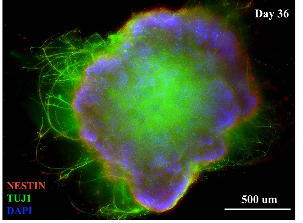
Human iPSC-Derived Cerebral Organoids are differentiated from hESC or iPSCs using Human iPSC-Derived Cerebral Organoid Differentiation Kit (Ca. No. RIPO-BWM001K). Cerebral organoids are three-dimensional in vitro models with a cellular composition and structural organization that is representative of the human cerebral regions. Organoids generated using Human iPSC-Derived Cerebral Organoid Differentiation Kit (Ca. No. RIPO-BWM001K) feature various types of neurons (including TH positive neurons) and glia cells (including OLIG2 and IBA1 positive cells).
产品特征(Product Specification)
The cerebral organoids are cryopreserved at 120 days under –80°C using the cerebral organoid cryopreservation medium. Cryopreservation and shipment will not cause deactivation of the cerebral organoids.
存储(Storage)
Upon arrival, if the organoids will not be used immediately, please store them under -80 °C. For thaw and recovery, please recover the organoids in the provided recovery medium with water bath for 48h. After recovery, please store the organoid in its maintenance medium under the correct incubation condition and medium changing process.
产品示意(Product Diagram)
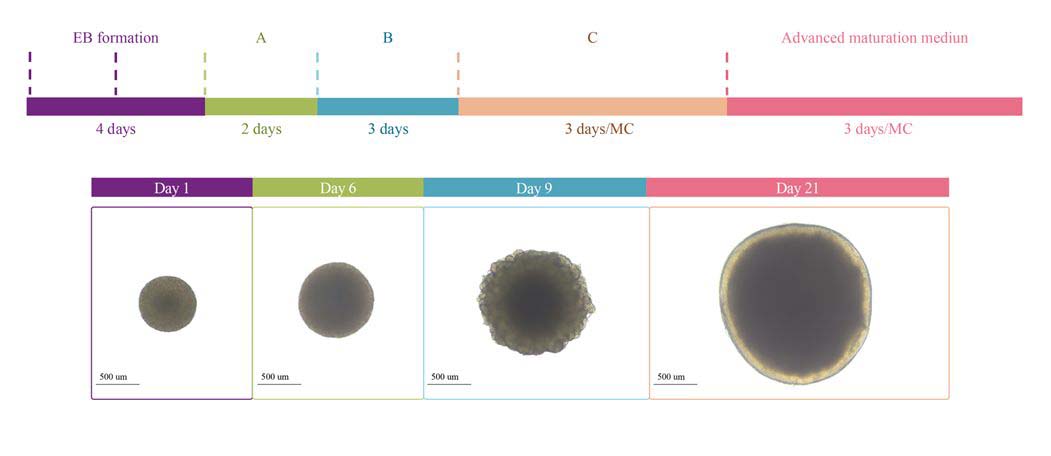
Protocol Diagram of cerebral organoid differentiation.
验证数据(Validation Data)
Orgnaoid Histology
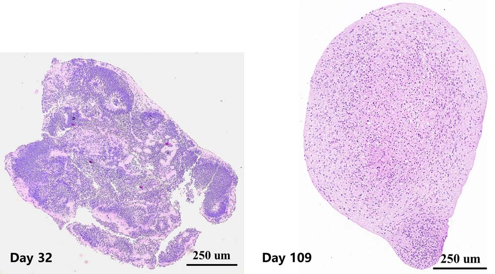
Left: Early-stage cerebral organoid show rosette-like structures (neural stem cells), which become smaller as organoids develop.
Right: Day 109 cerebral organoids show uniform morphology and show no dead core inside.
Marker Expression
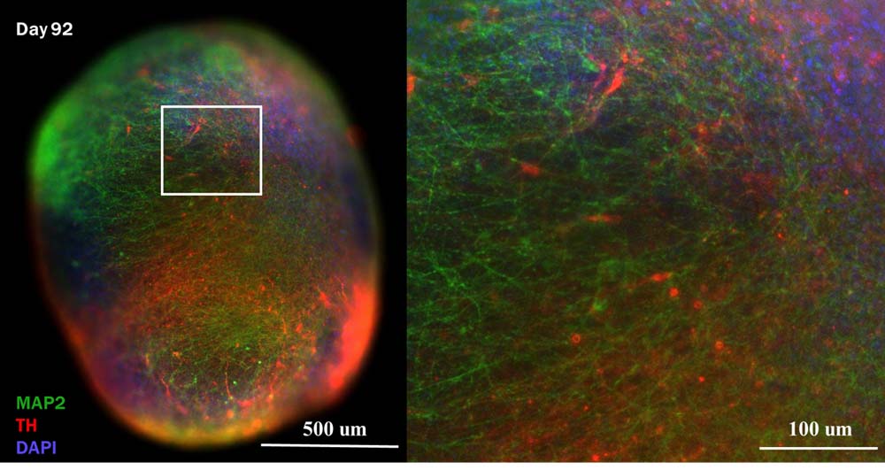
Presence of TH and MAP2 positive neurons in day 92 cerebral organoid.
TH: used as cell marker of dopaminergic neurons.
MAP2: mature neuron cell marker.
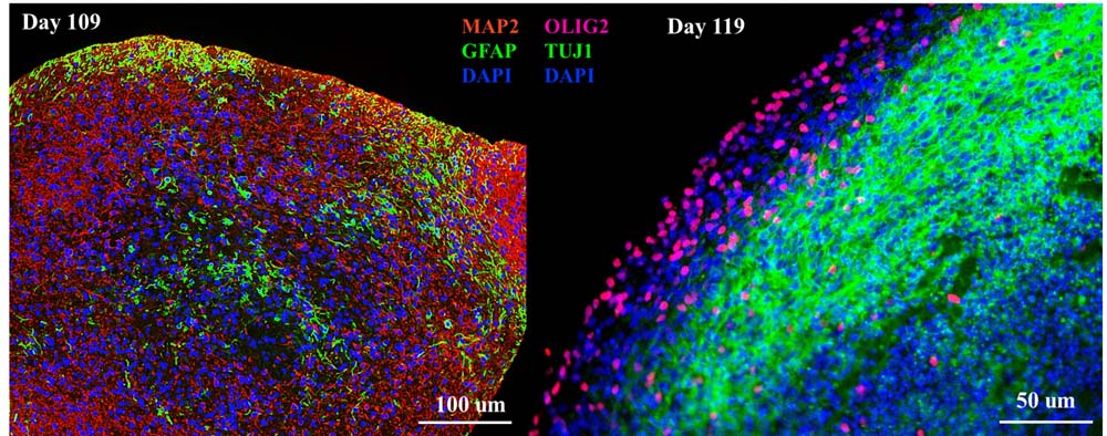
Left: Presence of GFAP positive cells at day 109 cultured cerebral organoid.
Right: Presence of OLIG2 positive cells at day 119 cultured cerebral organoid.
GFAP: marker for astrocyte.
OLIG2: marker for oligodendrocyte.
Presence of TH and IBA1 positive cell in day 147 cultured cerebral organoid.
IBA1: cell marker for microglia.
Organoid Activity
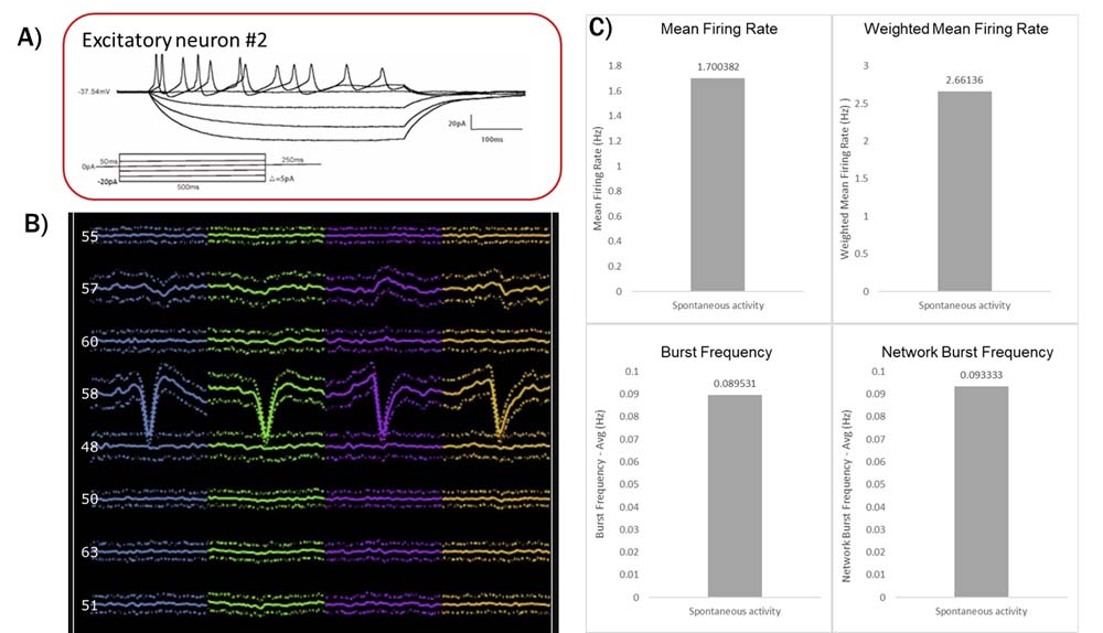
A: patch-clamp recording of excitatory neurons dissociated from cerebral organoids at day 102. Excitatory neuron show stable response to step injection currents.
B: Recording of cerebral organoid (day 60) using silicon probe. Neurons that show spontaneous activities have different waveforms.
C: MEA recording of cerebral organoid (day 86), spontaneous activities and bursting activities are recorded.
Organoid Application
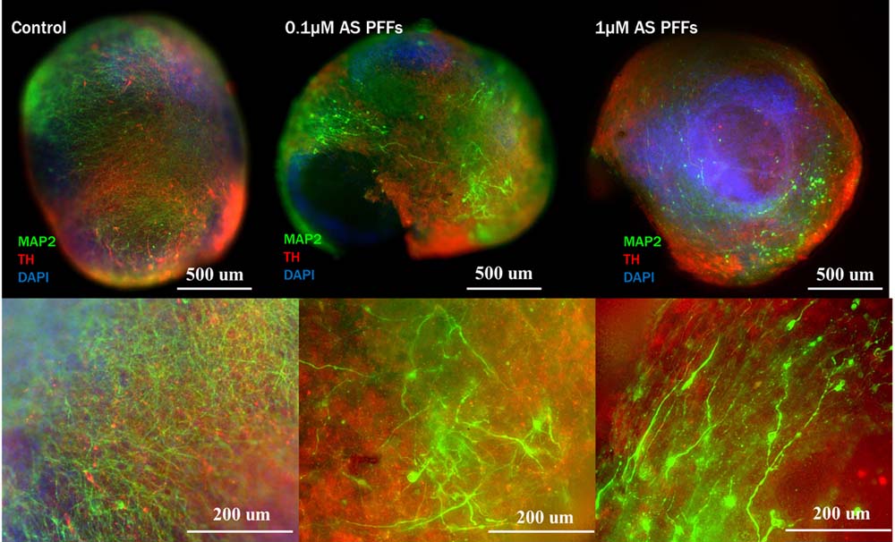
Using cerebral organoids for modelling Parkinson's Disease. Cerebral organoids are treated with Human Alpha-Synuclein Pre-formed Fibrils Protein (Ca. No. ALN-H51H4). Dopaminergic neurons and MAP2 positive neurons are damaged with both 0.1μM and 1μM Alpha-Synuclein Pre-formed Fibrils.

AAVs selection using cerebral organoids: Different capsid of AAVs were incubated with cerebral organoids. As the results, different affinities of each AAV to the cerebral organoid were observed.


 膜杰作
膜杰作 Star Staining


