产品描述(Product Details)
| Assay Type | Inhibition-TR-FRET |
| Analyte | Anti-TL1A Neutralizing Antibody |
| Format | 100T/500T |
| Reactivity | Human |
| Regulatory Status | RUO |
| Sensitivity | IC50=2.816nM |
| Standard Curve Range | 0.1302 nM-66.6667 nM |
| Assay Time | 1 hr |
| Suitable Sample Type | For screening assay of neutralizing antibodies binding to the human TL1A |
| Sample volume | 10 μL |
背景(Background)
TNF-like cytokine 1A (TL1A) and its receptors, death receptor 3 (DR3) and decoy receptor 3 (DcR3) are members of the TNF and TNF receptor superfamilies of proteins, respectively. Binding of APC-derived TL1A to lymphocytic DR3 provides co-stimulatory signals for activated lymphocytes. DR3 signaling affects not only the proliferative activity of and cytokine production by effector lymphocytes, but also critically influences the development and suppressive function of regulatory T-cells. Whereas, DcR3 restricts the function of the TL1A/DR3 complex: attenuating T-cell activation and downregulating the secretion of pro-inflammatory cytokines. Together with DR3 and DcR3, TL1A constitutes a cytokine system that actively interferes with the regulation of immune responses. The Human TL1A-DR3 Binding Kit (TR-FRET) can detect the binding of TL1A to DR3 in homogeneous system within 0.5-1 hours. it is high sensitive, short detection time and easy to use.
应用说明(Application)
The kit is developed for the binding of TL1A to the DR3.
It is for research use only.
存储(Storage)
1. Unopened kit should be stored at 2℃-8℃ upon receiving.
2. Find the expiration date on the outside packaging and do not use reagents past their expiration date.
3. The opened kit should be stored per components table. The shelf life is 30 days from the date of opening.
组分(Materials Provided)
| ID | Components | Size |
| FRT02-C01 | Human TL1A Protein Europium-chelate | 100 tests/500 tests |
| FRT02-C02 | FA Labeled Human DR3 Protein | 100 tests/500 tests |
| FRT02-C03 | Monoclonal Anti-TL1A Antibody, Human IgG1 | 20 μg/100 tests
100 μg/500 tests |
| FRT02-C04 | Sample Dilution Buffer | 10 mL/100tests & 500tests |
| FRT02-C05 | Detection Buffer | 10 mL/100tests & 500tests |
原理(Assay Principles)
This Human TL1A-DR3 inhibition kit (TR-FRET) is based on TR-FRET technology (Time-Resolved Fluorescence Resonance Energy Transfer). Use a mixture of biotinylated human TL1A and Europium-chelate labeled streptavidin as the donor and FA labeled human DR3 protein as the acceptor.
Your experiment will include 3 simple steps:
1) Mix the sample or Human Anti-TL1A Neutralizing Antibody in the kit with Human TL1A Protein Europium-chelate (Donor) and incubate at room temperature for 0.5 hours.
2) Add FA labeled Human DR3 Protein (Acceptor) and incubate at room temperature for at least 0.5 hours.
3) Use the TR-FRET module of a microplate reader to read the fluorescence signal at 665nm and 620nm. Calculate the Ratio based on the formula Ratio = "Signal 665 nm" / "Signal 620 nm" × 10^4. The Ratio value is negatively correlated with the inhibitors in the sample.
— When the sample does not contain the inhibitors of human TL1A binding to human DR3, the donor and acceptor are in close proximity because of the binding of human TL1A and FA labeled human DR3. The 620nm signal emitted by the donor under specific light source excitation is received by the acceptor, emitting a 665nm signal.
— When the sample contains the inhibitors of human TL1A binding to human DR3, the inhibitors prevent the binding between the donor and acceptor and thereby prevent FRET from occurring.
活性(Bioactivity)-TR-FRET Please refer to DS document for the assay protocol.

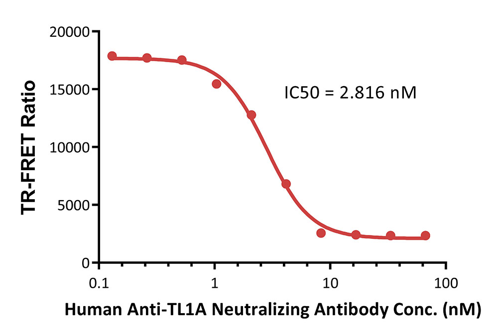
Inhibition of Europium-chelate labeled human TL1A: FA labeled human DR3 binding by Human Anti-TL1A Neutralizing Antibody
Premix serial dilutions of Human Anti-TL1A Neutralizing Antibody (1:2 serial dilution, from 10 μg/mL to 0.01953125 μg/mL (66.6667-0.1302 nM)) and Human TL1A Protein Europium-chelate and incubate at room temperature (20℃-25℃) for 0.5 hours. Then add FA Labeled Human DR3 Protein and incubate at room temperature (20℃-25℃) for 0.5 hours. Detection was performed with IC50 of 2.816nM. The assay was performed according to the above-described Datasheet (QC tested).
背景(Background):TL1A-DR3
The TL1A protein, belongs to the tumor necrosis factor (TNF) family and is a receptor for TNFRSF25 and TNFRSF6B. TL1A is involved in the activation of NF-κB and C-Jun pathways, which can be used as a regulator of mucosal immunity and participate in the immune pathway of inflammatory bowel disease (IBD) pathogenesis.


 膜杰作
膜杰作 Star Staining


