登录 | 注册    关注公众号  
微信公众号
搜索
 >  Kits>protein L >RES-A026

resDetect™ Protein L ELISA Kit

For research use only.

产品参数(Product Specifications)

Assay TypeSandwich-ELISA
AnalyteProtein L
Format96T(8×12 strips)
Regulatory StatusRUO
Sensitivity<50pg/mL
Standard Curve Range50 pg/mL-3200 pg/mL
Assay Time2 hr
Suitable Sample TypeFor the quantitative determination of recombinant protein L
Sample volume50uL

产品概述(Product Overview)

resDetect™ Protein L ELISA Kit is based on the ELISA sandwich method and is used to measure natural or structurally conserved recombinant forms of Protein L, as well as recombinant forms of Protein L with significant structural differences from natural Protein A, such as MabSelect™ VL, in bioprocess manufacturing applications. It is designed to provide a reliable solution to aid in optimal purification process development and routine quality control of in-process streams and final products. It can also be used as a universal detection tool for the quantitative determination of Protein L.

存储(Storage)

1. Unopened kit should be stored at 2℃-8℃ upon receiving.

2. Find the expiration date on the outside packaging and do not use reagents past their expiration date.

3. The opened kit should be stored per components table. The shelf life is 30 days from the date of opening.

组分(Materials Provided)

IDComponentsSize
RES026-C01Pre-Coated Anti-Protein L Antibody Microplate1 plate(8×12 strips)
RES026-C02Recombinant Protein L Standard (1μg/mL)100μL
RES026-C03Biotin-Anti-Protein L Antibody150uL
RES026-C04Streptavidin-HRP10μg
RES026-C0510×Sample Dilution Buffer15mL
RES026-C06Denaturation Buffer15mL
RES026-C0720×Washing Buffer30mL
RES026-C08Antibody Dilution Buffer15mL
RES026-C09Streptavidin-HRP Dilution Buffer15mL
RES026-C10Substrate Solution12mL
RES026-C11Stop Solution8mL

质量管理控制体系(QMS)

  1. 质量管理体系(ISO, GMP)
  2. 质量优势
  3. 质控流程
 

典型数据-Typical Data Please refer to DS document for the assay protocol.

protein L TYPICAL DATA

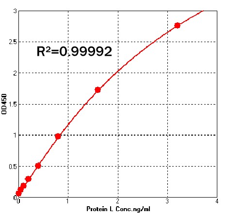
Detection of Recombinant Protein L by sandwich-ELISA Assay. Immobilized Anti- Protein L Antibody can bind Recombinant Protein L . Detection was performed using Biotin-Anti- Protein L Antibody with sensitivity of 50 pg/mL. For each experiment, a standard curve needs to be set for each micro-plate, and the specific OD value may vary depending on different laboratories, testers, or equipments. The following example data is for reference only.


验证(Validation)

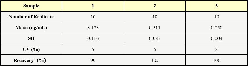
批内差异(Intra-Assay Statistics)

Three samples of known concentration were tested ten times on one plate to assess intra-assay precision.

protein L INTRA-ASSAY STATISTICS

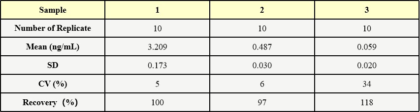
批间差异(Inter-Assay Statistics)

Three samples of known concentration were tested in three separate assays to assess inter-assay precision.
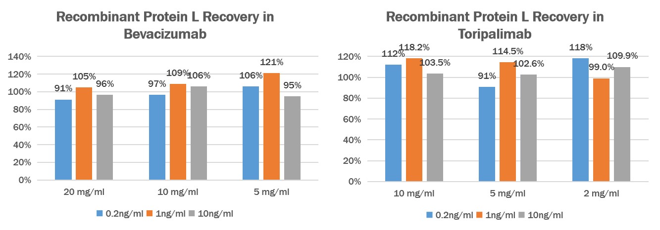
protein L INTER-ASSAY STATISTICS

回收率(Recovery)

Add different concentrations of Protein L (0.2ng/mL、1ng/mL、10ng/mL) to different concentrations of Human IgG1 (Bevacizumab) (20mg/mL、10mg/mL、5mg/mL) or Human IgG4 (Toripalimab) (10mg/mL、5mg/mL、2mg/mL), then dilute the antibodies to a reasonable range, then test and calculated the concentration of protein L to give the recovery rate.

protein L RECOVERY

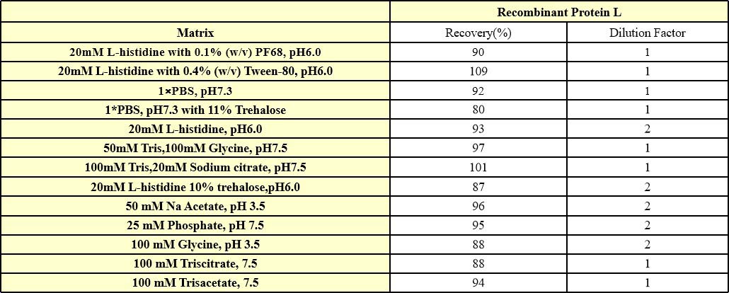
干扰效应(Interference effect)

We have conducted interference effect test about frequently-used buffers, they have excellent buffer compatibility. For specific buffers, it is recommended that you verify recovery to determine the minimum dilution ratio.
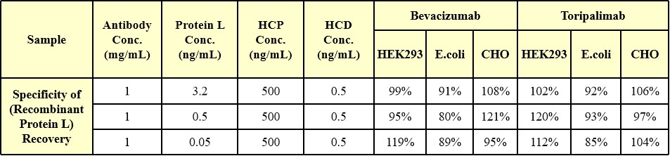
protein L INTERFERENCE EFFECT

专属性(Specificity)

Host cell protein (HCP 500 ng/mL) and host cell DNA (HCD 0.5 ng/mL) of HEK293, E.coli or CHO systems were added to human IgG1 (Bevacizumab, 1mg/mL) and human IgG4 (Toripalimab, 1mg/mL), respectively, which were higher than the usual quality standard limit. Then high, medium, and low concentrations of Protein L were added, respectively, and the ratio of Protein L recovery in the Protein L added samples without HCP and HCD was added as the specificity verification index. verification index.

protein L SPECIFICITY

 
评论(0)
 
ACRO质量管理体系


背景(Background):protein L

Protein L was isolated from the surface of bacterial species Peptostreptococcus magnus and was found to bind Ig(IgG,IgM,IgA,IgE and IgD) through L chain interaction, from which the name was suggested. Despite this wide-ranging binding capability with respect to Ig classes, Protein L is not a universal immunoglobilin-binding protein. Binding of Protein L to immunoglobulins is restricted to those containing kappa light chains (i.e., k chain of the VL domain). In humans and mice, kappa (k) light chains predominate. The remaining immunoglobulins have lambda (l) light chains. The recombinant protein contains four immunoglobulin (Ig) binding domains (Bdomains) of the native protein. Besides antibody, protein L is also suitable for binding of a wide range of antibody fragments such as Fabs, single-chain variable fragments (scFv), and domain antibodies (Dabs).


前沿进展

Esketamine mitigates systemic inflammation via modulating phenotypic transformation of monocytes in patients undergoing thoracic surgery
Shen, Yan, Zhang et al
Life Sci (2025)
Abstract: To assess esketamine's anti-inflammatory effects during thoracic surgery and its modulation of immune responses.In a randomized trial, 64 of 73 patients undergoing thoracic surgery were allocated into the Control (not receiving esketamine) or the ES-KTM group (intraoperative esketamine infusion). Blood routine tests were conducted one day before (T0) and one day after the surgery (T3). Plasma levels of tumor necrosis factor-α (TNF-α) and interleukine-10 (IL-10) were analyzed by ELISA, and cell surface markers including CD14, CD16, CD163, CD40, CX3CR1, CD206 were tested by cytometry at the entry to the surgical room (T1) and the skin closure (T2). For the in vitro study, esketamine at 10 μM was employed to treat the lipopolysaccharide (LPS) stimulated macrophage cell line-Raw264.7, and its effects were tested by cytometry and RNA sequencing analysis.Esketamine application reduces the count of neutrophils and monocytes, neutrophil to lymphocyte ratio (NLR), platelet to lymphocyte ratio (PLR) and systemic inflammatory index (SII), and enhances the lymphocyte counting and lymphocyte to monocyte ratio (LMR). Then, esketamine application decreases the plasma TNF-α levels, while maintaining the IL-10 level in comparison with the Control group. Additionally, esketamine reduced the proportion of intermediate monocytes, downregulates the expressions of CD16, CD40 and CX3CR1, while upregulates the CD206 expression. Finally, in the in-vitro study, esketamine inhibits the M1 pro-inflammatory markers in LPS-challenged macrophages, and downregulates multiple immune-related pathways.Esketamine mitigates surgery-triggered inflammation by suppressing monocyte/macrophage proinflammatory activity and TNF-α release, offering dual anesthetic and immunomodulatory benefits.Copyright © 2025. Published by Elsevier Inc.
The Causal Relationship Between Immune Cells and Neuropathic Pain: A Two-Sample Mendelian Randomization Study Based on Genome-Wide Association Analysis
Li, Liu
J Pain Res (2025) 18, 1515-1523
Abstract: Increasing evidence indicates that various types of immune cells are associated with different forms of neuropathic pain (NP). However, the causal relationships among these associations remain unclear. To elucidate the causal relationships between immune cells and NP, we conducted a two-sample Mendelian randomization (MR) analysis.The exposure and outcome Genome-wide association analysis (GWAS) data used in this study were obtained from open-access databases. This study employed a two-sample MR analysis to evaluate the causal relationships between 731 immune cell traits and four types of NP, including postherpetic neuralgia (PHN), trigeminal neuralgia (TN), diabetic peripheral neuropathy (DPN), and drug-induced peripheral neuropathy (DIPN).The relative count of CD39+ CD4+ %T cells was positively associated with TN, while the mean fluorescence intensity (MFI) of CD20 on IgD+ CD38br (B cell) and forward scatter area (FSC-A) on myeloid dendritic cells (DCs) were negatively associated with TN. Additionally, the relative count of CD8br NKT %lymphocytes was positively associated with PHN, and the MFI of HLA DR on CD33br HLA DR+ CD14 (myeloid cells) was negatively associated with PHN. The MFI of CD4 on activated and secreting T regulatory (Treg) cells was positively associated with DPN. Furthermore, the relative count of B cell % CD3- lymphocytes was negatively associated with DIPN.This MR study, using genetic data from individuals of European descent, provides evidence supporting the causal relationships between several specific immune cell phenotypes and various NP subtypes.© 2025 Li and Liu.
Genetically Supported Causality Between Immune Cells Traits and Low Back Pain: A Bi-Directional Two-Sample Mendelian Randomization Study
Wang, Zhu, Liu et al
J Pain Res (2025) 18, 1577-1585
Abstract: Prior studies have suggested that immune cells play a crucial role in Low Back Pain (LBP). We employed a bi-directional Mendelian randomization (MR) study to investigate the causal relationship of immune cells with the risk of LBP.Single Nucleotide Polymorphisms (SNPs) that had a significant genetic association with immune cells were used as instrumental variables (IVs). The inverse variance weighted (IVW) method was used as the primary approach for MR analyses. To assess the robustness, sensitivity analyses were further performed using MR-Egger and MR-PRESSO.The MR analysis revealed a causal relationship between six types of immune cells and LBP (P < 0.05), including CD4 Treg AC (OR, 0.925; 95% CI, 0.878-0.974; P = 0.003), CD19 on CD20- CD38- (OR, 0.938; 95% CI, 0.898-0.979; P = 0.003), CD4 on HLA DR+ CD4+ (OR, 0.947; 95% CI, 0.909-0.987; P = 0.010), CD25 on CD39+ CD4+ (OR, 0.954; 95% CI = 0.922-0.988; P = 0.008), CD14 on CD33br HLA DR+ CD14dim (OR, 0.950; 95% CI = 0.916-0.985; P = 0.006), and CD4RA on TD CD4+ (OR, 1.030; 95% CI, 1.012-1.048; P = 0.001). Reverse MR analysis found no evidence of potential causal effects of genetically predicted LBP on the six types of immune cells.This study has demonstrated a close genetic connection between immune cells and LBP, providing valuable insights for future clinical research.© 2025 Wang et al.
Causal Relationship Between Immune Cells and Venous Thromboembolism: A Bidirectional Two-Sample Mendelian Randomization Study
Su, Li, Wen et al
Vasc Health Risk Manag (2025) 21, 181-195
Abstract: Manny evidence indicates that numerous immune cells are linked to the onset and progression of VTE, though the causal relationship remains unclear. To determine the association between immune cells and VTE, we performed a bidirectional two-sample Mendelian randomization (MR) study.A comprehensive MR analysis was conducted to ascertain the causal relationship between immune cell signatures and VTE. Leveraging publicly available genetic data, we examined the causal associations between 731 immune cell signatures and the risk of VTE. The analysis encompassed four types of immune signatures, namely median fluorescence intensities, relative cell counts, absolute cell counts, and morphological parameters. We employed the two-sample MR analysis, used the inverse variance-weighted (IVW) approach as the primary analytical method. Rigorous sensitivity analyses were employed to validate the robustness, heterogeneity, and presence of horizontal pleiotropy in the results. Furthermore, the reverse MR analysis was implemented to confirm the existence of reverse causal relationships.Eighteen immune cell signatures were found to have nominally significant associations with VTE according to the IVW method. The level of CD14 expression on CD14+ CD16+ monocytes (OR 0.95) and ten other phenotypes were identified as protective factors against VTE. Conversely, the percentage of HLA DR+ T cells among lymphocytes (OR 1.03) and six other phenotypes were identified as risk factors associated with an increased likelihood of VTE. The expression level of CX3CR1 on CD14- CD16+ monocytes showed a potential bidirectional causal relationship.Our study identified 18 types of immune cell signatures that could impact VTE development, offering novel insights for future mechanistic and clinical studies in this field. Further studies to prospectively validate our findings are needed.© 2025 Su et al.
Showing 1-4 of 30964 papers.
Powered by BizGenius
 
 
货号/价格
文档
联系电话:
+86 400-682-2521(全国)
010-53681107(北京)
021-50850665(上海)
运输方式
订单邮箱:
order.cn@acrobiosystems.com
技术支持邮箱:
tech.cn@acrobiosystems.com
前沿进展
点击查看详细

消息提示

请输入您的联系方式,再点击提交!

确定