登录 | 注册    关注公众号  
微信公众号
搜索
 >  Protein>DR3 >DR3-H82E3

Biotinylated Human DR3 / TNFRSF25 Protein, His,Avitag™ (MALS verified)

分子别名(Synonym)

TNFRSF25,DR3,APO3,DDR3,TNFRSF12,WSL,WSL1

表达区间及表达系统(Source)

Biotinylated Human DR3 Protein, His,Avitag (DR3-H82E3) is expressed from human 293 cells (HEK293). It contains AA Gln 25 - Gln 199 (Accession # Q93038-1).

Request for sequence

蛋白结构(Molecular Characterization)

DR3 Structure

This protein carries a polyhistidine tag at the C-terminus, followed by an Avi tag (Avitag™).

The protein has a calculated MW of 22.6 kDa. The protein migrates as 30-35 kDa when calibrated against Star Ribbon Pre-stained Protein Marker under reducing (R) condition (SDS-PAGE) due to glycosylation.

标记(Labeling)

Biotinylation of this product is performed using Avitag™ technology. Briefly, the single lysine residue in the Avitag is enzymatically labeled with biotin.

蛋白标记度(Protein Ratio)

Passed as determined by the HABA assay / binding ELISA.

内毒素(Endotoxin)

Less than 1.0 EU per μg by the LAL method.

纯度(Purity)

>90% as determined by SDS-PAGE.

>90% as determined by SEC-MALS.

制剂(Formulation)

Lyophilized from 0.22 μm filtered solution in PBS, pH7.4 with trehalose as protectant.

Contact us for customized product form or formulation.

重构方法(Reconstitution)

Please see Certificate of Analysis for specific instructions.

For best performance, we strongly recommend you to follow the reconstitution protocol provided in the CoA.

存储(Storage)

For long term storage, the product should be stored at lyophilized state at -20°C or lower.

Please avoid repeated freeze-thaw cycles.

This product is stable after storage at:

  1. -20°C to -70°C for 12 months in lyophilized state;
  2. -70°C for 3 months under sterile conditions after reconstitution.

质量管理控制体系(QMS)

  1. 质量管理体系(ISO, GMP)
  2. 质量优势
  3. 质控流程
 

电泳(SDS-PAGE)

DR3 SDS-PAGE

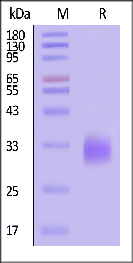
Biotinylated Human DR3 Protein, His,Avitag on SDS-PAGE under reducing (R) condition. The gel was stained with Coomassie Blue. The purity of the protein is greater than 90% (With Star Ribbon Pre-stained Protein Marker).

SEC-MALS

DR3 SEC-MALS

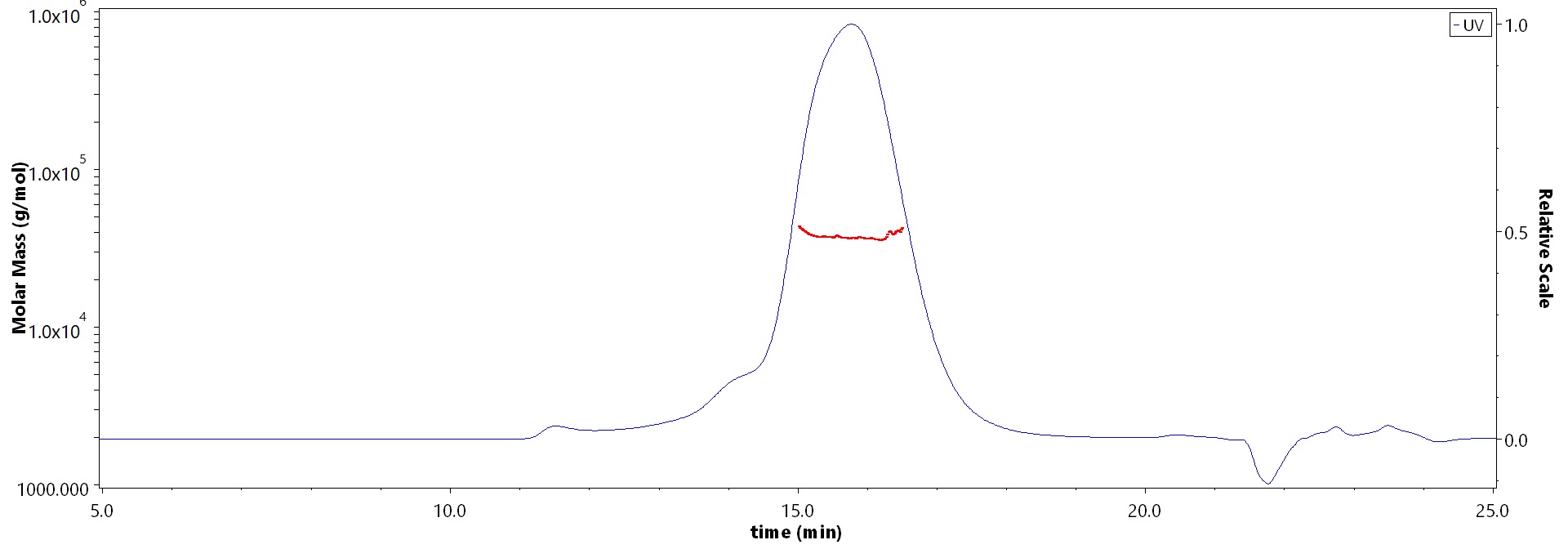
The purity of Biotinylated Human DR3 Protein, His,Avitag (Cat. No. DR3-H82E3) is more than 90% and the molecular weight of this protein is around 30-45 kDa verified by SEC-MALS.

Report


活性(Bioactivity)-ELISA

DR3 ELISA

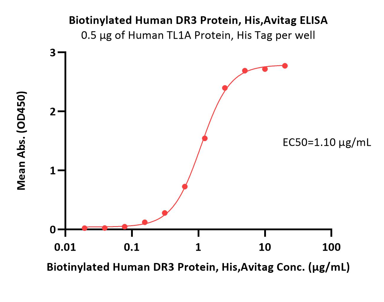
Immobilized Human TL1A Protein, His Tag (Cat. No. TLA-H5244) at 5 μg/mL (100 μL/well) can bind Biotinylated Human DR3 Protein, His,Avitag (Cat. No. DR3-H82E3) with a linear range of 0.02-2.5 μg/mL (QC tested).

Protocol


活性(Bioactivity)-SPR

DR3 SPR

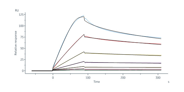
Biotinylated Human DR3 Protein, His,Avitag (Cat. No. DR3-H82E3) captured on Biotin CAP-Series S Sensor Chip can bind Human TL1A, His Tag (Cat. No. TLA-H5243) with an affinity constant of 1.75 nM as determined in a SPR assay (Biacore 8K) (Routinely tested).

Protocol

 
评论(0)
 
ACRO质量管理体系


背景(Background)

Tumor necrosis factor receptor superfamily member 25 (TNFRSF25) is also known as Apo-3, Death receptor 3 (DDR3 or DR3), Apoptosis-inducing receptor AIR, Apoptosis-mediating receptor TRAMP, Lymphocyte-associated receptor of death, Apo-3, which is a member of the TNF-receptor superfamily. TNFRSF25 is a homodimer protein, which can Interact strongly via the death domains with TNFRSF1 and TRADD to activate at least two distinct signaling cascades, apoptosis and NF-kappa-B signaling. TNFRSF25 is receptor for TNFSF12 / APO3L / TWEAK.


前沿进展

Design, Synthesis and Anti-Influenza Virus Activity of 4-Tert-Butyl-N-(3-Oxo-1-Thia-4-Azaspiro[4.5]Dec-4-yl)Benzamide Derivatives That Target Hemagglutinin-Mediated Fusion
Çınar, Alikadıoğlu, Soylu-Eter et al
Drug Dev Res (2025) 86 (2), e70080
Abstract: Hemagglutinin (HA) is a viral glycoprotein that mediates influenza virus entry into the host cell and is considered a relevant viral target. We here report the identification of a class of 4-tert-butylphenyl-substituted spirothiazolidinones as HA-mediated fusion inhibitors with specific activity against influenza A/H3N2 virus. The novel spirocyclic compounds were achieved by using one-pot cyclocondensation method and the chemical structures were characterized by IR, 1H NMR, 13C NMR, and elemental analysis. Compound 2c, bearing methyl substitutions at positions 2- and 8- of the spiro ring displayed an EC50 value against influenza A/H3N2 virus of 1.3 μM and an antiviral selectivity index of 30. The fusion-inhibiting effect of compound 2c was revealed in the polykaryon assay which is based on cell-cell fusion when influenza virus H3 HA-transfected cells are exposed to low pH. Computer-aided docking was performed to predict the possible binding pocket in the H3 HA trimer. Resistance data and in silico studies indicated that compound 2c has an overlapping binding pocket in the stem region of H3 HA with the known fusion inhibitors TBHQ and arbidol.© 2025 The Author(s). Drug Development Research published by Wiley Periodicals LLC.
High soluble expression and characterization of human GalNAc transferase T2 and T11 in Escherichia coli
Wang, Zhang, Shi et al
Protein Expr Purif (2025) 231, 106712
Abstract: The efficient expression of soluble glycosyltransferases from mammalian sources in Escherichia coli (E. coli) remains a significant challenge, often resulting in misfolding and the formation of inclusion bodies. In this study, we investigated strategies to enhance the solubility and catalytic activity of human GalNAc-T2 and GalNAc-T11, two O-glycosyltransferases involved in O-glycosylation of glycoproteins. We found that fusion with maltose-binding protein (MBP) and cellulase catalytic domain (Cel-CD), which led to majority of the fusion proteins being soluble, could increase the solubility of the recombinant proteins. Enzyme activity assays revealed that the fusion glycosyltransferase exhibited significantly higher catalytic efficiency than non-fused enzymes. In addition, the influence of GalNAc-T11 lectin domain on substrate specificity was also determined. The presence of lectin domain had no influence on the recognition of specific substrate and the specific activity of GalNAc-T11. This work offers an efficient approach for the large-scale production of human glycosyltransferases with enhanced bioactivity, highlighting its potential for glycosylation engineering of glycoprotein drugs.Copyright © 2025. Published by Elsevier Inc.
Real-world evidence provides clinical insights into tissue-agnostic therapeutic approvals
Sledge, Yoshino, Xiu et al
Nat Commun (2025) 16 (1), 2646
Abstract: The US Food and Drug Administration approves tissue-agnostic therapies to target tumor biomarkers regardless of tumor type. In light of the growing number of such approvals in recent years, a better understanding of their relative clinical benefit across cancer types is required. To address this need, we analyzed tissue-agnostic indications (TMB-High, MSI-High/MMRd, BRAFV600E mutations, and NTRK and RET fusions) in a database of 295,316 molecularly-profiled tumor samples with associated clinical outcomes data. Here, we show that 21.5% of tumors harbored at least one of the tissue-agnostic indications investigated, including 5.4% lacking a cancer-specific indication. Our analysis reveals poor uptake of targeted therapies for rare NTRK fusions, significant differences in pembrolizumab-associated outcomes across tumor types for TMB-High and MSI-High/MMRd, as well as clinical benefits in tumor types and drugs of the same class not investigated in the pivotal clinical trials. These results demonstrate that treatment effects are not necessarily tissue-agnostic, and suggest possible expansion of therapeutic avenues for a given tissue-agnostic indication.© 2025. The Author(s).
Histopathology of C Cells and Medullary Thyroid Carcinoma
Cameselle-Teijeiro, Sobrinho-Simões
Recent Results Cancer Res (2025) 223, 9-50
Abstract: C cells are the neuroendocrine cell component of the thyroid gland that embryologically arise from the pharyngeal endoderm. Normal C cells are concentrated in the upper two-thirds of both lateral lobes, appear singly or in small groups dispersed in, among or peripherally to the follicles, and are involved in the production of calcitonin. Reactive C-cell hyperplasia should be differentiated from proliferation of atypical C cells (neoplastic C-cell hyperplasia) which is considered an intraepithelial neoplasia of C cells/medullary carcinoma in situ, a precursor lesion associated to familial medullary thyroid carcinoma (MTC). MTC typically exhibits a lobular and/or trabecular growth pattern with amyloid deposits; however, due to its great histological variability, immunohistochemical positivity for calcitonin, carcinoembryonic antigen, calcitonin-gene-related peptide, insulinoma-associated protein 1, and/or other markers is necessary to confirm diagnosis. Investigation of germline RET proto-oncogene mutation is mandatory to identify familial MTC. Somatic RET mutations or fusions as well as RAS mutations in cytological and/or biopsy samples may represent therapeutic targets. Mixed medullary and follicular-derived cell carcinoma is a heterogeneous group of tumors which needs to be distinguished from collision tumors.© 2025. The Author(s), under exclusive license to Springer Nature Switzerland AG.
Showing 1-4 of 35217 papers.
Powered by BizGenius
 
 
货号/价格
产品推荐
文档
联系电话:
+86 400-682-2521(全国)
010-53681107(北京)
021-50850665(上海)
运输方式
订单邮箱:
order.cn@acrobiosystems.com
技术支持邮箱:
tech.cn@acrobiosystems.com
DR3靶点信息
英文全称:Tumor necrosis factor receptor superfamily member 25
中文全称:肿瘤坏死因子受体超家族成员25
种类:Homo sapiens
上市药物数量:0详情
临床药物数量:0详情
最高研发阶段:临床前
查看更多信息
前沿进展
点击查看详细

消息提示

请输入您的联系方式,再点击提交!

确定