登录 | 注册    关注公众号  
微信公众号
搜索
 >  Protein>CCR2 >CC2-H52P3

Human CCR2 Full Length Protein (VLP)

分子别名(Synonym)

C-C chemokine receptor type 2,C-C CKR-2,CC-CKR-2,CC-CKR-2CKR2B,CCR2,CCR-2,CCR2A,CCR2B,CD192 antigen,CD192,chemokine (C-C motif) receptor 2,CKR2,CKR2A,CMKBR2,CMKBR2MGC111760,FLJ78302,MCP-1 receptor,MCP-1-R,MCP-1-RMGC103828,MGC168006,Monocyte chemoattractant protein 1 receptor,monocyte chemotactic protein 1 receptor

表达区间及表达系统(Source)

Human CCR2 Full Length Protein (VLP) (CC2-H52P3) is expressed from human 293 cells (HEK293). It contains AA Met 1 - Leu 360 (Accession # P41597-2).

Predicted N-terminus: Met 1

蛋白结构(Molecular Characterization)

Virus-like particles(VLPs) are formed by self-assembly of envelop/capsid proteins from viruses. Membrane Proteins can be constituted in-situ with VLPs produced from HEK293 cell cultures. These VLPs concentrate conformationally intact membrane proteins directly on the cell surface and produce soluble, high-concentration proteins perfect for immunization and antibody screening.

The VLPs provide the display of properly folded membrane proteins in their native cellular membrane in a compact size of 100~300 nm diameter (similar to the size of most viruses) making it optimal targets for dendritic cells in vivo and surface attachment for phage display.

内毒素(Endotoxin)

Less than 1.0 EU per μg by the LAL method.

制剂(Formulation)

The VLPs are highly immunogenic, so the immunization strategy should be optimized (antigen dose, regimen and adjuvant).

Supplied as 0.2 μm filtered solution in PBS, pH7.4 with glycerol as protectant.

Contact us for customized product form or formulation.

运输(Shipping)

This product is supplied and shipped with dry ice, please inquire the shipping cost.

存储(Storage)

Please avoid repeated freeze-thaw cycles.

This product is stable after storage at:

  1. The product MUST be stored at -70°C or lower upon receipt;
  2. -70°C for 12 months under sterile conditions.

质量管理控制体系(QMS)

  1. 质量管理体系(ISO, GMP)
  2. 质量优势
  3. 质控流程
*The isotype control of empty/mock VLP (Cat. No. VLP-N5213) is sold separately and not included in protein, you can follow this link for product information.
 

活性(Bioactivity)-ELISA

CCR2 ELISA

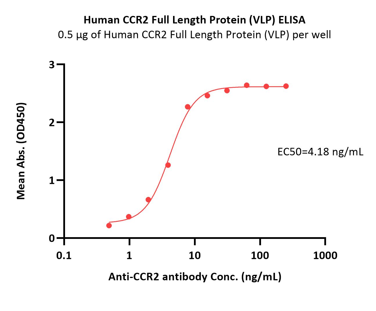
Immobilized Human CCR2 Full Length Protein (VLP) (Cat. No. CC2-H52P3) at 5 μg/mL (100 μL/well) can bind Anti-CCR2 antibody with a linear range of 0.5-8 ng/mL (QC tested).

Protocol


均一性(Identity)-DLS

CCR2 DLS

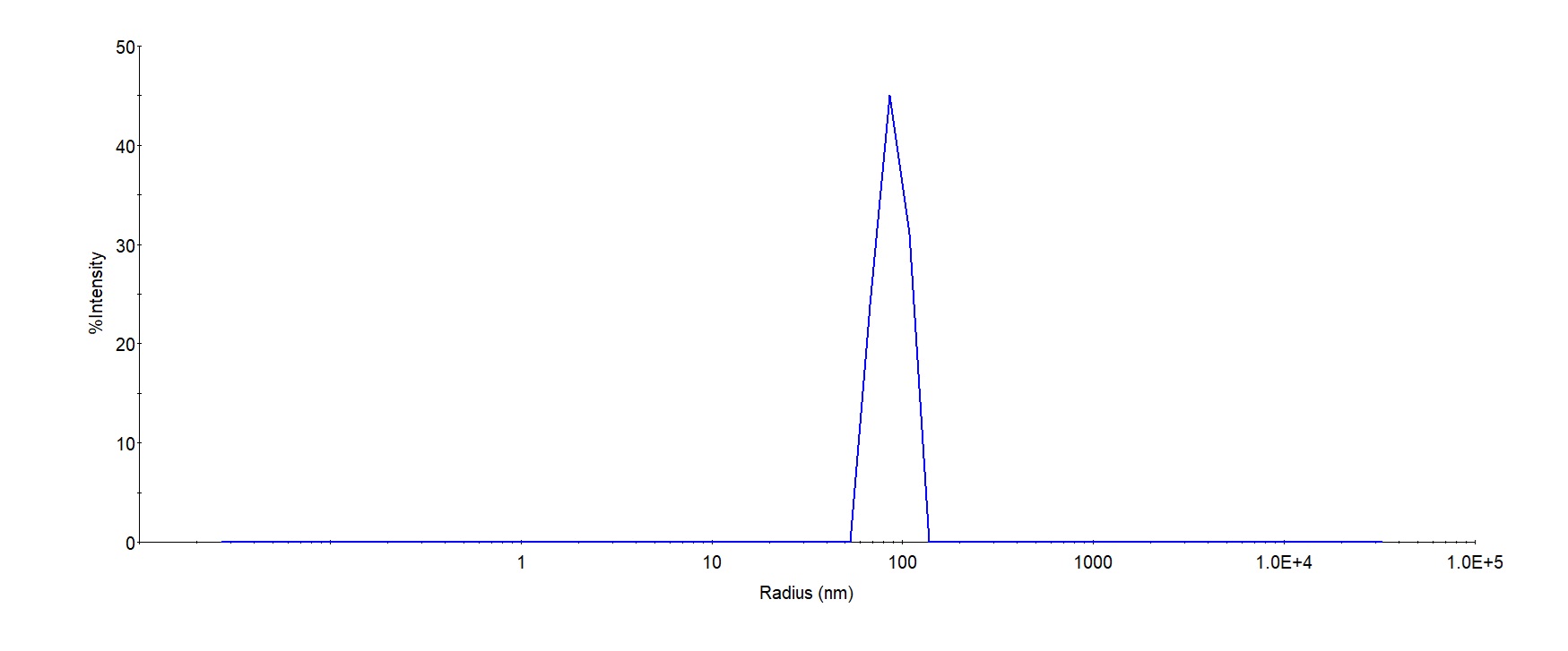
The mean peak Radius of VLP is 80-120 with more than 95% intensity as determined by dynamic light scattering (DLS).

 
评论(0)
 
ACRO质量管理体系


背景(Background)

The protein encoded by this gene is a receptor for monocyte chemoattractant protein-1, a chemokine which specifically mediates monocyte chemotaxis. Monocyte chemoattractant protein-1 is involved in monocyte infiltration in inflammatory diseases such as rheumatoid arthritis as well as in the inflammatory response against tumors. The encoded protein mediates agonist-dependent calcium mobilization and inhibition of adenylyl cyclase. This protein can also be a coreceptor with CD4 for HIV-1 infection. This gene is located in the chemokine receptor gene cluster region of chromosome 3.


前沿进展

Endothelial-targeting miR-145 micelles restore barrier function and exhibit atheroprotective effects
Ashraf, Huang, Choroomi et al
Nanoscale Horiz (2025)
Abstract: Atherosclerosis remains the leading cause of death worldwide and is characterized by the accumulation of plaque beneath the endothelium. MicroRNA-145-5p (miR-145), which is downregulated in atherosclerosis, has been shown to mitigate plaque development by promoting the contractile vascular smooth muscle cell (VSMC) phenotype. Previously, our lab found that miR-145 micelles conjugated with MCP-1 peptides were able to inhibit atherosclerosis by targeting diseased VSMC via C-C chemokine receptor 2 (CCR2). Diseased endothelial cells similarly express CCR2; however, the impact of miR-145 micelles on endothelial cell function has not been explored. Thus, in this study, the in vitro therapeutic effects of miR-145 micelles in modulating the endothelium during atherosclerosis are evaluated. To that end, the MCP-1 peptide density on the micelle surface was first optimized for activated endothelial cell binding, followed by loading miR-145 into micelles with the optimal MCP-1 ratio. Following characterization, miR-145 micelle treatment of activated endothelial cells resulted in efficient miR-145 transfection, upregulation of atheroprotective genes, and suppression of atherogenic genes. Furthermore, the treatment enhanced the integrity of endothelial tight junctions and reduced monocyte migration. This work establishes miR-145 micelles as an effective nanotherapeutic for restoring endothelial cell health in cardiovascular disease for the first time.
Human umbilical cord mesenchymal stem cell-derived microvesicles alleviate pulmonary fibrosis by inhibiting monocyte‒macrophage migration through ERK1/2 signaling-mediated suppression of CCL2 expression
Liang, Li, Wu et al
Stem Cell Res Ther (2025) 16 (1), 145
Abstract: Pulmonary fibrosis (PF) is a disease with high morbidity and mortality rates, but effective treatment options are extremely limited. Mesenchymal stem cells (MSCs) and their derivatives show promise as potential therapeutics for PF. However, the underlying mechanisms responsible for these beneficial effects remain poorly understood. The objective of this study was to elucidate the specific mechanism through which microvesicles derived from human umbilical cord MSCs (MSC-MVs) alleviate PF.The effects of MSC-MVs on PF in bleomycin (BLM)-induced mice were assessed via histological staining, flow cytometry, and enzyme-linked immunosorbent assays (ELISAs). The potential therapeutic target was identified via RNA sequencing (RNA-seq) analysis, followed by validation via real-time quantitative polymerase chain reaction (RT‒qPCR), ELISAs, scratch testing, and western blotting (WB).MSC-MVs significantly attenuated collagen fiber deposition and downregulated the expression of extracellular matrix components in the lungs of the BLM-induced mice. Moreover, this treatment substantially ameliorated lung inflammation by reducing the monocyte‒macrophage ratio and the TNF-α and IL-6 levels. Further analyses revealed that MSC-MVs inhibited the classic chemotactic CCL2/CCR2 axis of monocyte‒macrophages, leading to reduced recruitment of monocytes‒macrophages to the lungs, which decreased lung inflammation and prevented fibrotic progression. Both in vitro and in vivo findings demonstrated that MSC-MVs suppressed ERK1/2 phosphorylation followed by decreased CCL2 production to modulate monocyte-macrophage migration.Our findings demonstrate that the protective effect of MSC-MVs against BLM-induced lung toxicity was achieved through the inhibition of the ERK1/2 signaling pathway, leading to the suppression of CCL2 expression and subsequent modulation of monocyte-macrophage migration, thereby establishing a theoretical basis for the effect of MSC-MVs in PF.© 2025. The Author(s).
Neoadjuvant BMS-813160, nivolumab, gemcitabine and nab-paclitaxel for patients with pancreatic cancer
Grierson, Wolf, Suresh et al
Clin Cancer Res (2025)
Abstract: Targeting tumor-associated macrophages through C-C chemokine receptor type 2 (CCRs) in pancreatic ductal adenocarcinoma (PDAC) improves the efficacy of chemotherapy and restores T cell immunity in preclinical models.We conducted a phase I/II single institution study (NCT03496662) combining chemotherapy gemcitabine and nab-paclitaxel (GnP), CCR2/5 inhibitor BMS-813160 and nivolumab for four 28-day cycles for patients with borderline resectable (BR) or locally advanced (LA) PDAC. The recommended phase 2 dose (RP2D) of BMS-813160 was established in the 3+3 design. Primary endpoints were safety and objective response rate (ORR). Secondary endpoints included resection rate, median progression-free survival (mPFS) and overall survival (mOS).8 patients were treated with GnP alone (control arm) and 31 patients (29 response evaluable) were treated at RP2D. No grade 3/4 toxicities attributed to nivolumab or BMS-813160 were identified. After 4 cycles of study treatment (N=26), ORR was 35.7% and 16.7% among BR- and LA-PDAC patients respectively, compared to 0% of control patients. 78.6% BR- and 16.7% of LA-PDAC patients who completed study treatment underwent surgical resection. For intent-to-treat analyses, BR-PDAC patients had a mPFS and mOS of 14.6 and 20.4 months respectively; and for LA-PDAC patients, were 14.7 and 17 months, respectively. Biomarker analyses showed decreased intratumoral monocytes, macrophages, enhanced T cell proliferation and effector gene expression.Neoadjuvant BMS-813160/nivolumab/GnP was well tolerated and appears to achieve comparable ORR and resectability to historical data, however with prolonged PFS and OS in LA-PDAC patients, warranting a larger phase II study with a more efficacious CCR2-targeted therapeutic.
Alveolar epithelial type 2 cell specific loss of IGFBP2 activates inflammation in COVID-19
Pujadas, Chin, Sankpal et al
Respir Res (2025) 26 (1), 111
Abstract: The coronavirus disease 2019 (COVID-19) global pandemic is caused by severe acute respiratory syndrome coronavirus 2 (SARS-CoV-2). However, our understanding of SARS-CoV-2-induced inflammation in alveolar epithelial cells remains very limited. The contributions of intracellular insulin-like growth factor binding protein-2 (IGFBP2) to SARS-CoV-2 pathogenesis are also unclear. In this study, we have uncovered a critical role for IGFBP2, specifically in alveolar epithelial type 2 cells (AEC2), in the immunopathogenesis of COVID-19. Using bulk RNA sequencing, we show that IGFBP2 mRNA expression is significantly downregulated in primary AEC2 cells isolated from fibrotic lung regions from patients with COVID-19-acute respiratory distress syndrome (ARDS) compared to those with idiopathic pulmonary fibrosis (IPF) alone or IPF with a history of COVID-19. Using multicolor immunohistochemistry, we demonstrated that IGFBP2 and its selective ligands IGF1 and IGF2 were significantly reduced in AEC2 cells from patients with COVID-ARDS, IPF alone, or IPF with COVID history than in those from age-matched donor controls. Further, we demonstrated that lentiviral expression of Igfbp2 significantly reduced mRNA expression of proinflammatory cytokines-Tnf-α, Il1β, Il6, Stat3, Stat6 and chemokine receptors-Ccr2 and Ccr5-in mouse lung epithelial cells challenged with SARS-CoV-2 spike protein injury (S2; 500 ng/mL). Finally, we demonstrated higher levels of cytokines-TNF-α; IL-6 and chemokine receptor-CCR5 in AEC2 cells from COVID-ARDS patients compared to the IPF alone and the IPF with COVID history patients. Altogether, these data suggest that anti-inflammatory properties of IGFBP2 in AEC2 cells and its localized delivery may serve as potential therapeutic strategy for patients with COVID-19.© 2025. The Author(s).
Showing 1-4 of 6068 papers.
Powered by BizGenius
 
 
货号/价格
文档
联系电话:
+86 400-682-2521(全国)
010-53681107(北京)
021-50850665(上海)
运输方式
订单邮箱:
order.cn@acrobiosystems.com
技术支持邮箱:
tech.cn@acrobiosystems.com
前沿进展
点击查看详细
相关产品
View All CCR2

消息提示

请输入您的联系方式,再点击提交!

确定