[Mechanism of electroacupuncture in improving cartilage tissue injury in rats with knee osteoarthritis by regulating miR-335-5p expression]Zhu, Yan, Chen
et alZhen Ci Yan Jiu (2025) 50 (3), 310-318
Abstract: To observe the protective effect of electroacupuncture (EA) on the knee joint cartilage tissue of rats with knee osteoarthritis (KOA), so as to explore the potential mechanism of miR-335-5p involved in the treatment of EA for KOA.A total of 42 adult male Sprague-Dawley (SD) rats were randomly divided into blank, model and EA groups, with 14 rats in each group. The model was established by injecting a sodium glutamate iodoacetate solution (80 μg/μL) into the right joint cavity. On the 15th day after the model establishment, rats in the EA group received EA intervention at "Yanglingquan" (GB34) and "Xuehai" (SP10) on the right side, and the needles were retained for 15 minutes. The intervention was carried out every other day for a total of two weeks. After the two-week intervention, the paw withdrawal mechanical threshold (PWMT) and paw withdrawal thermal latency (PWTL) of the rats were detected;a gait analyzer was used to detect the gait adaptability changes induced by pain;the pathological changes of the knee joint cartilage tissue were observed after hematoxylin-eosin (HE) staining;qPCR and Western blot were used to detect the expression levels of miR-335-5p, Dickkopf related protein 1 (DKK-1), β-catenin, and matrix metalloproteinase 13 (MMP-13) mRNAs and proteins in the cartilage tissue respectively;immunofluorescence staining was used to observe the positive fluorescence expression of β-catenin in the right knee joint cartilage tissue.In the model group, HE staining showed a large area of chondrocyte necrosis, with the dissolution and disappearance of cell nuclei, accompanied by a macrophages-predominant inflammatory cell infiltration;cartilage surface defects in local areas, necrosis of trabecular bone and proliferation of fibrous tissue in local areas, and new capillary formation in the proliferative areas were also observed. In the EA group, pathological manifestations such as inflammatory cell infiltration and fibrous tissue proliferation were significantly alleviated. Compared with the blank group, the PWMT, PWTL, the support duration, footprint area, and average ground pressure of the right hind paw, and the mRNA and protein expressions of DKK-1 in the right knee joint cartilage tissue were decreased (P<0.001) in the model group, while the expressions of miR-335-5p, mRNA and protein expressions of β-catenin and MMP-13, and the fluorescence intensity of β-catenin in the right knee joint cartilage tissue were increased (P<0.001, P<0.01). All the above indicators in the EA group were all reversed (P<0.001, P<0.05, P<0.01) in comparison with the model group.EA can significantly improve the pain symptoms of KOA rats, promote the recovery of motor function, and improve the pathological morphology of the knee joint cartilage tissue. This may be related to regulating the expression of miR-355-5p and Wnt/β-catenin pathway, as well as inhibiting the release of cartilage extracellular matrix-related degrading enzymes.
LRP5 enhances glioma cell proliferation by modulating the MAPK/p53/cdc2 pathwayFeng, Jin, Pan
et alInt J Med Sci (2025) 22 (4), 990-1001
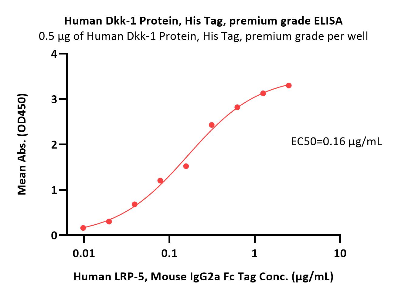
Abstract: Background: Glioma is a malignant neoplasm with generally poor prognosis and the treatment options and effective drugs are very limited. LRP5, a member of the low-density lipoprotein receptor (LDLR) gene family, has been reported to regulate the progression of various cancers such as gastric and colorectal cancer. However, the function of LRP5 in glioma has not been elucidated. The objective of this study is to explore the influence of LRP5 in glioma cell proliferation and its potential molecular mechanisms. Methods: LRP5 expression in glioma was assessed through bioinformatics analysis, and validation was conducted using clinical glioma tissues. Glioma cell lines with reduced LRP5 expression were established through RNA interference. A series of experiments such as cell proliferation assay, flow cytometry analysis, and Western blotting were used to determine the role of LRP5 in glioma cell proliferation, cell cycle progression, and the underlying mechanisms. Results: LRP5 was found to be upregulated in glioma tissues and exhibited significant variations across various subtypes of glioblastoma (GBM). When differentiating between normal individuals and glioma patients, the area under the receiver operating characteristic curve (ROC) for LRP5 was determined to be 0.981. Downregulating the expression of LRP5 in glioma cells can weaken their proliferative ability and reduce the number of cell colonies. There were more cells arrested in the G2/M phase of the cell cycle. The protein levels of phospho-p53 (p-p53), p21Cip1, and phospho-cdc2 (p-cdc2) were elevated. Moreover, LRP5 down-regulation suppressed the phosphorylation of the mitogen-activated protein kinase (MAPK) family members, JNK and p38 MAPK. Consistent results with those mentioned above can be achieved by using an LRP5 antagonist named DKK-1. Conclusion: This research has identified that LRP5 may promote glioma proliferation by influencing the G2/M transition and the activation of the MAPK/p53/cdc2 pathways, suggesting its value as a potential molecular target for glioma diagnosis and treatment.© The author(s).
Study on the mechanism of Wnt/β-catenin pathway mediated by pterostilbene to reduce cerebral ischemia-reperfusion injuryJin, Fu, Guo
et alBiomol Biomed (2025)
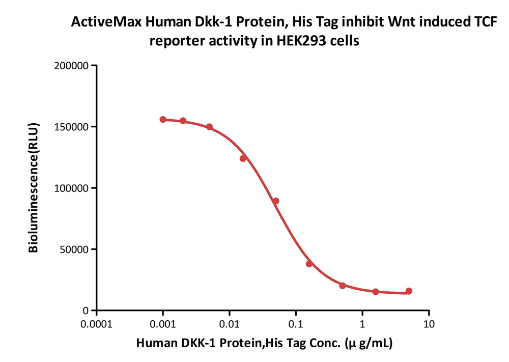
Abstract: Cerebral ischemia-reperfusion injury (CIRI) is the primary cause of damage following ischemic stroke, with ferroptosis serving as a key pathophysiological factor in CIRI. Pterostilbene (PTE) has been shown to reduce cerebral ischemic injury, but whether its mechanism of action involves ferroptosis remains unclear. In this study, an in vitro model of mouse hippocampal neuron (HT22) cell injury and an in vivo mouse CIRI model were established. Treatments included PTE, the ferroptosis activator Erastin, and the Wnt signaling pathway inhibitor (Dkk-1). Cell damage was assessed using flow cytometry, MTT assay, lactate dehydrogenase (LDH) release assay, and Calcein-AM/PI staining. Oxidative stress and ferroptosis in cells and tissues were evaluated using biochemical kits and fluorescence staining. Additionally, histopathological staining was performed to assess brain tissue damage, while qRT-PCR and Western blot analyses were used to measure ferroptosis-related factors and Wnt/β-catenin pathway-related proteins in both cells and tissues. HT22 cells subjected to injury exhibited decreased viability and increased cell death (P < 0.05). Similarly, CIRI mice demonstrated pronounced cerebral infarction and neuronal damage. Ferroptosis, characterized by elevated levels of iron ions, lipid peroxides (ROS and MDA), and reduced antioxidant enzymes (GSH and GPX4), was significantly increased in both cells and tissues (P < 0.05). Correspondingly, ferroptosis-related protein levels were elevated (P < 0.05), while Wnt/β-catenin pathway-related protein levels were significantly decreased (P < 0.05). Treatment with Erastin and Dkk-1 exacerbated neuronal damage, intensified ferroptosis, and inhibited the Wnt/β-catenin pathway. Conversely, PTE treatment activated the Wnt/β-catenin pathway, reduced ferroptosis, and improved neuronal damage. Specifically, PTE upregulated the Wnt/β-catenin pathway, decreased peroxide accumulation, and antagonized ferroptosis, ultimately mitigating CIRI. These findings suggest that PTE protects against CIRI by modulating the Wnt/β-catenin pathway and alleviating ferroptosis-induced damage.
Current Landscape of Molecular Biomarkers in Gastroesophageal Tumors and Potential Strategies for Co-Expression PatternsKorpan, Puhr, Berger
et alCancers (Basel) (2025) 17 (3)
Abstract: The treatment of metastasized gastroesophageal adenocarcinoma largely depends on molecular profiling based on immunohistochemical procedures. Therefore, the examination of HER2, PD-L1, and dMMR/MSI is recommended by the majority of clinical practice guidelines, as positive expression leads to different treatment approaches. Data from large phase-III trials and consequent approvals in various countries enable physicians to offer their patients several therapy options including immunotherapy, targeted therapy, or both combined with chemotherapy. The introduction of novel therapeutic targets such as CLDN18.2 leads to a more complex decision-making process as a significant number of patients show positive results for the co-expression of other biomarkers besides CLDN18.2. The aim of this review is to summarize the current biomarker landscape of patients with metastatic gastroesophageal tumors, its direct clinical impact on daily decision-making, and to evaluate current findings on biomarker co-expression. Furthermore, possible treatment strategies with multiple biomarker expression are discussed.


 膜杰作
膜杰作 Star Staining


