登录 | 注册    关注公众号  
微信公众号
搜索
 >  Cell>NKp46 >SCJUR-STF130

Human NKp46 (Luc) Jurkat Reporter Cell Development Service

For research use only.

  1. Genetically modified cell lines best reflect MOA (Mechanism of Action)
  2. Higher activity and larger assay window for robust and reproducible cell-based bioassay
  3. Comprehensive application data to support assay development and validation
  4. Full tracible record, stringent quality control and validated cell passage stability
  5. Parental cell line legally obtained from internationally recognized cell resource bank and commercially licensed
  6. Global commercial license assistance whenever regulatory filing is required

描述(Description)

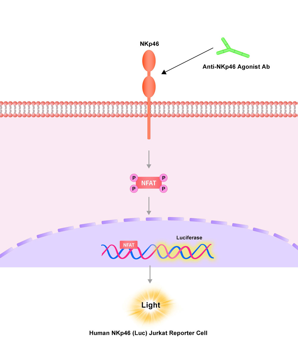
The Human NKp46 (Luc) Jurkat Reporter Cell was engineered to not only express the NFAT response element driving luciferase expressing systems, but also express the receptor full length human NKp46 (Gene ID: 9437). When cocultured with anti-human Nkp46 agonist antibody, the interaction of agonist antibody and NKp46 on the surface of Human NKp46 (Luc) Jurkat Reporter Cell results in NFAT-mediated luminescence.

应用说明(Application)

• Screen for anti-human NKp46 agonist antibody.

NKp46 Assay Principles

生长特性(Growth Properties)

Suspension

筛选标记(Selection Marker)

Puromycin (5 μg/mL) + Hygromycin (20 μg/mL)

培养基(Complete Growth Medium)

RPMI-1640 + 10% FBS

冻存液(Freeze Medium)

Serum-free cell cryopreservation medium

装量(Quantity)

1 vial contains at least 5×10^6 cells in 1 mL serum-free cryopreservation medium

存储(Storage)

Frozen in liquid nitrogen.

支原体检测(Mycoplasma Testing)

Negative

无菌检测(Sterility Testing)

Negative

使用说明(Instructions for Use)

See data sheet for detailed culturing and assay protocol.

质量管理控制体系(QMS)

  1. 质量管理体系(ISO, GMP)
  2. 质量优势
  3. 质控流程
 

Receptor Assay

NKp46 FACS

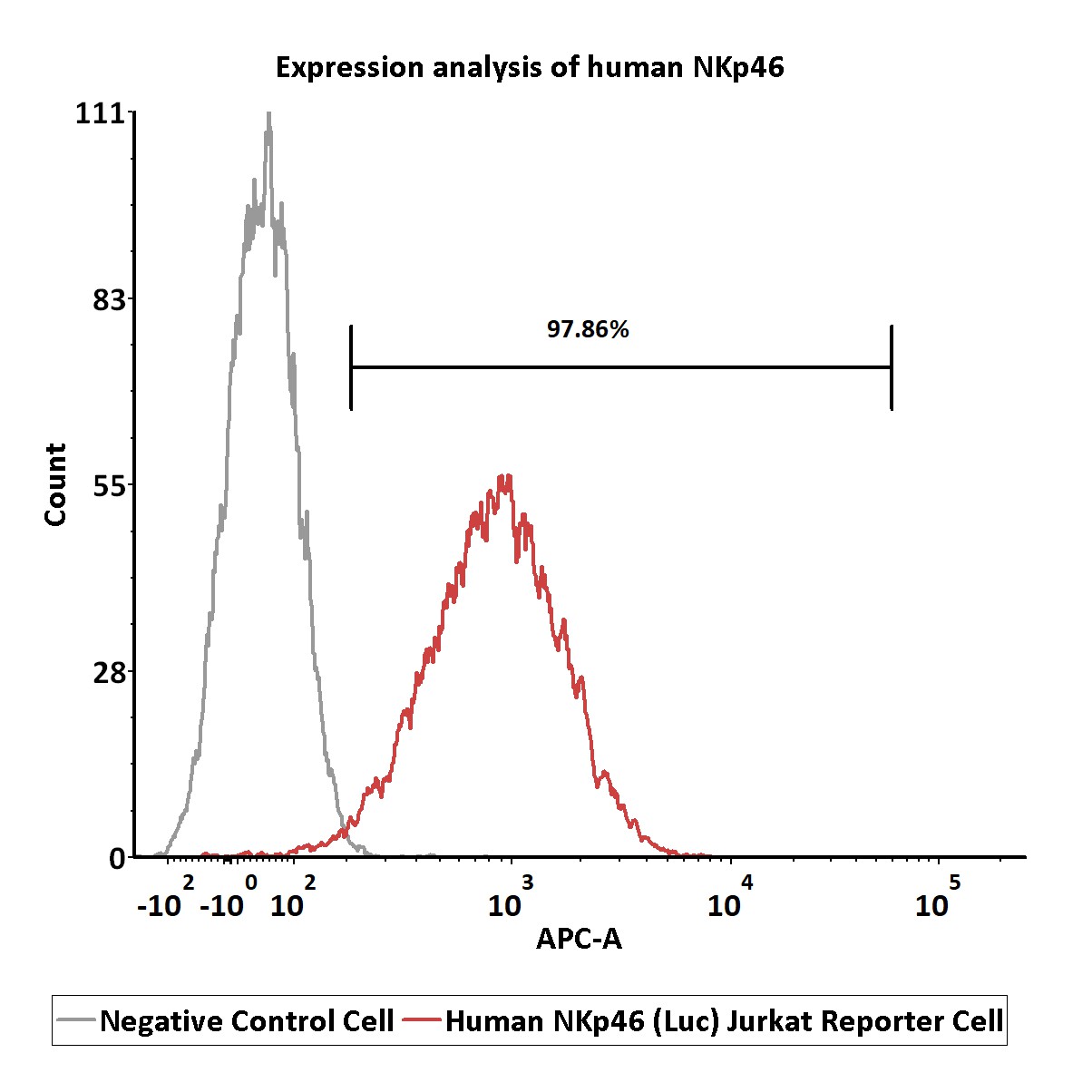
Expression analysis of human NKp46 on Human NKp46 (Luc) Jurkat Reporter Cell by FACS.
Cell surface staining was performed on Human NKp46 (Luc) Jurkat Reporter Cell or negative control cell using APC- labeled Anti-human NKp46 antibody.

Protocol


Application

NKp46 APPLICATION

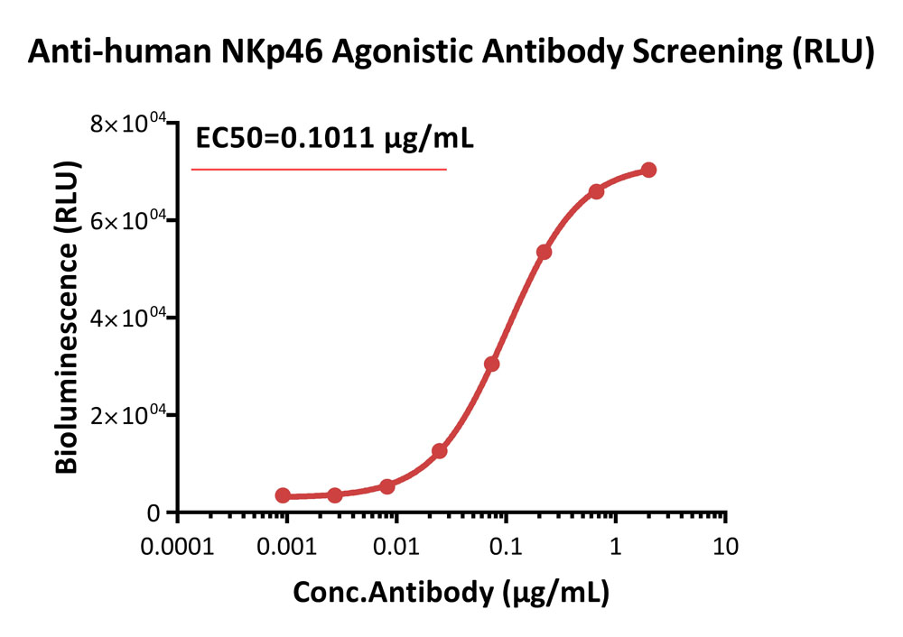
Agonistic activity analysis of anti-human NKp46 antibody (RLU).
This reporter cell was incubated with serial dilutions of anti-human NKp46 antibody. The EC50 of anti-human NKp46 antibody was approximately 0.1011 μg/mL.

Protocol

NKp46 APPLICATION

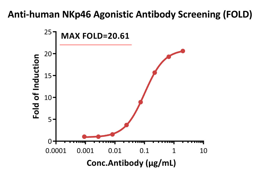
Agonistic activity analysis of anti-human NKp46 antibody (FOLD).
This reporter cell was incubated with serial dilutions of anti-human NKp46 antibody. The max induction fold was approximately 20.61.

Protocol


Passage Stability

NKp46 PASSAGE

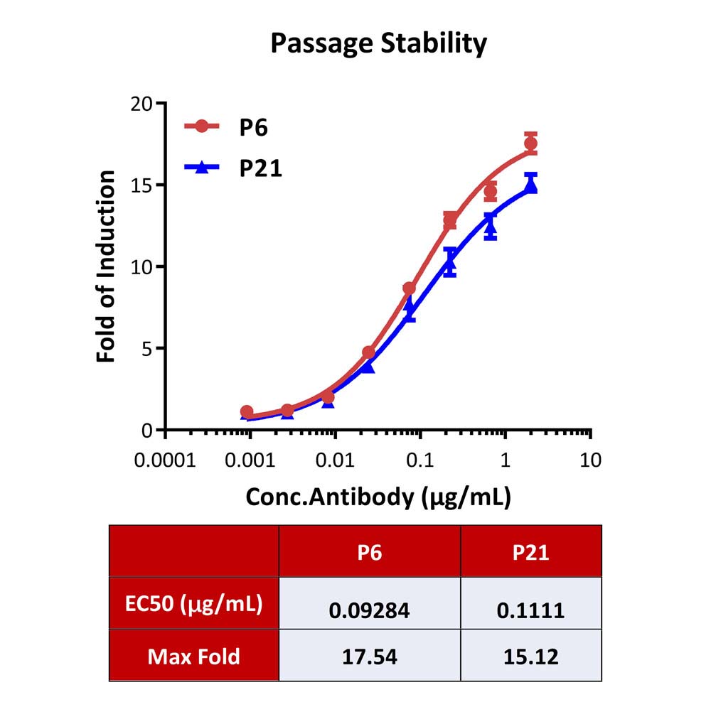
Passage stability analysis by Signaling Bioassay.
The continuously growing Human NKp46 (Luc) Jurkat Reporter Cell was stimulated with serial dilutions of anti-human NKp46 antibody. Anti-human NKp46 antibody stimulated response demonstrates passage stabilization (fold induction and EC50) across passage 6-21.

Protocol

如有相关细胞池需求请联系我们

 
评论(0)
 
ACRO质量管理体系


背景(Background)

Natural cytotoxicity triggering receptor 1 (NCR1) is also known as Natural killer cell p46-related protein (NK-p46), Lymphocyte antigen 94 homolog (LY94), CD antigen CD335, which belongs to the natural cytotoxicity receptor (NCR) family. NCR1 contains two Ig-like (immunoglobulin-like) domains. NCR1 interacts with CD247 and FCER1G. NCR1 / CD335 may contribute to the increased efficiency of activated natural killer (NK) cells to mediate tumor cell lysis.

Limited Use&License Disclosure

BY USE OF THIS PRODUCT, RESEARCHER AGREES TO BE BOUND BY THE FOLLOWING TERMS OF LIMITED USE OF THIS CELL LINE PRODUCT.

  1. If the researcher is not willing to accept the terms of limited use of this cell line product, and the product is unused, ACRO will accept return of the unused product.
  2. Researchers may use this product for research use only, no commercial use is allowed. "Commercial use" means any and all uses of this product and derivatives by a party for profit or other consideration and may include but is not limited to use in: (1) product manufacture; and (2) to provide a service, information or data; and/or resale of the product or its derivatives, whether or not such product or derivatives are resold for use in research.
  3. This cell line is neither intended for any animal or human therapeutic purposes nor for any direct human in vivo use . You have no right to share, modify, transfer, distribute, sell, sublicense, or otherwise make the cell line available for use to other researchers, laboratories, research institutions, hospitals, universities, or service organizations.
  4. ACROBIOSYSTEMS MAKES NO WARRANTIES OR REPRESENTATIONS OF ANY KIND, EITHER EXPRESSED OR IMPLIED, WITH RESPECT TO THE SUITABILITY OF THE CELL LINE FOR ANY PARTICULAR USE.
  5. ACROBIOSYSTEMS ACCEPTS NO LIABILITY IN CONNECTION WITH THE HANDLING OR USE OF THE CELL LINE.
  6. Modifications of the cell line, transfer to a third party, or commercial use of the cell line may require a separate license and additional fees. Please contact order.cn@acrobiosystems.com for further details.


前沿进展

Rosuvastatin restores liver tissue-resident NK cell activation in aged mice by improving mitochondrial function
Amer, Salhab, Safadi
Biomed Pharmacother (2025) 186, 118000
Abstract: Aging has an impact on Natural Killer (NK) cells surveillance against tumors and infections. Our study aims to assess the aging effects on metabolic and mitochondrial markers influencing NK cell activity.C57BL/6 J mice aged 12, 24, 48, and 72 weeks were used. Liver injury serum and histological markers, pro-inflammatory cytokines [IL-1β, IL-2, IL-6] and chemoattractant markers [CCL2, CXCL8] were assessed. Moreover, cholesterol metabolic markers [HMG-CoA synthetase, HMG-CoA reductase, mevalonate kinase], mitochondrial biogenesis [PGC1α] and functional gene markers [TFAM, HSPA9, Seahorse, apoptosis] in liver trNK cells, were assessed by RT-PCR. Senescence [p16, p21], exhaustion [PD-1, TIGIT, LAG3], activation [CD107a, NKp46], and chemokine receptor [CCR2, CXCR1] markers were assessed in trNK cells using flow cytometry. Liver trNK cells of aged mice were treated with Rosuvastatin [10μM] for 12 h.Data showed a linear increase in liver injury markers, pro-inflammatory and chemotaxis along aging. These results were associated with reductions in liver trNK cell counts and activations with a noticeable decrease in their chemoattractant receptor expressions. TrNK cells of aged mice exhibited elevated markers of senescence and exhaustion with a gradual increase in cholesterol accumulation. Mitochondrial biogenesis and functional gene markers showed a decrease in their expressions in aged mice while ameliorated following rosuvastatin treatment. Results were correlated with a decrease in cholesterol metabolism and restoring their NK cell activity.Our study demonstrates age-related cholesterol accumulation in trNK cells correlated with senescence and functional impairment. Rosuvastatin is suggested to boost, rejuvenate and recover NK cell functionality.Copyright © 2025. Published by Elsevier Masson SAS.
Enteric tuft cell inflammasome activation drives NKp46+ILC3 IL22 via PGD2 and inhibits Salmonella
Churchill, Pandeya, Bauer et al
J Exp Med (2025) 222 (6)
Abstract: To distinguish pathogens from commensals, the intestinal epithelium employs cytosolic innate immune sensors. Activation of the NAIP-NLRC4 inflammasome initiates extrusion of infected intestinal epithelial cells (IEC) upon cytosolic bacterial sensing. We previously reported that activation of the inflammasome in tuft cells, which are primarily known for their role in parasitic infections, leads to the release of prostaglandin D2 (PGD2). We observe that NAIP-NLRC4 inflammasome activation in tuft cells leads to an antibacterial response with increased IL-22 and antimicrobial protein levels within the small intestine, which is dependent on PGD2 signaling. A NKp46+ subset of ILC3 expresses the PGD2 receptor CRTH2 and is the source of the increased IL-22. Inflammasome activation in tuft cells also leads to better control of Salmonella Typhimurium in the distal small intestine. However, tuft cells in the cecum and colon are dispensable for antibacterial immunity. These data support that intestinal tuft cells can also induce antibacterial responses, possibly in a tissue-specific manner.© 2025 Churchill et al.
Gene-modified NK cells expressing CD64 and preloaded with HIV-specific BNAbs target autologous HIV-1-infected CD4+ T cells by ADCC
Tomescu, Ochoa-Ortiz, Lu et al
J Immunol (2025) 214 (2), 253-264
Abstract: Natural killer (NK) cells can efficiently mediate antibody-dependent cellular cytotoxicity (ADCC) of antibody coated target cells via the low-affinity Fc-receptor, CD16, but cannot retain antibodies over time. To increase antibody retention and facilitate targeted ADCC, we genetically modified human NK cells with the high-affinity Fc receptor, CD64, so that we could preload them with HIV-specific broadly neutralizing antibodies (BNAbs) and enhance their capacity to target HIV-infected cells via ADCC. Purified NK cells from the peripheral blood of control donors or persons living with HIV were activated with interleukin (IL)-2/IL-15/IL-21 cytokines and transduced with a lentivirus encoding CD64. High levels of CD64 surface expression were maintained for multiple weeks on NK cells and CD64-transduced NK cells were phenotypically similar to control NK cells with strong expression of CD56, CD16, NKG2A, NKp46, CD69, HLA-DR, CD38, and CD57. CD64-transduced NK cells exhibited significantly greater capacity to bind HIV-specific BNAbs in short-term antibody binding assay as well as retain the BNAbs over time (1-wk antibody retention assay) compared with control NK cells only expressing CD16. BNAb-preloaded CD64-transduced NK cells showed a significantly enhanced capacity to mediate ADCC against autologous HIV-1-infected CD4+ primary T cells in both a short-term 4 h degranulation assay as well as a 24 h HIV p24 HIV elimination assay when compared with control NK cells. A chimeric CD64 enhanced NK cell strategy (NuKEs [NK Enhancement Strategy]) retaining bound HIV-specific BNAbs represents a novel autologous primary NK cell immunotherapy strategy against HIV through targeted ADCC.© The Author(s) 2025. Published by Oxford University Press on behalf of The American Association of Immunologists. All rights reserved. For commercial re-use, please contact reprints@oup.com for reprints and translation rights for reprints. All other permissions can be obtained through our RightsLink service via the Permissions link on the article page on our site—for further information please contact journals.permissions@oup.com.
Blood biomarker dynamics in people with relapsing multiple sclerosis treated with cladribine tablets: results of the 2-year MAGNIFY-MS study
Wiendl, Barkhof, Montalban et al
Front Immunol (2025) 16, 1512189
Abstract: Cladribine tablets (CladT) represent an effective immune reconstitution therapy, administered in short treatment courses over two consecutive years. To better understand the amplitude of immune changes, we performed a comprehensive analysis during the 2-year study period for the entire MAGNIFY-MS population (N=270). In addition to lymphocyte kinetics, we studied intracellular cytokines serum proteins, and their associations with clinical outcomes. To put these changes into perspective, we analyzed transcriptional changes in T and B cells and associated biological pathways before and after each treatment course with CladT.Immunophenotyping and transcriptomics were performed at regular visits with major differences reported between baseline (BL) and after each yearly treatment course. Assessments included: lymphocyte dynamics, RNA sequencing (B and T cells), intracellular cytokines, serum proteins (immunoglobulins [IgG and IgM], and serum neurofilament light chain [sNfL]). Clinical measures included: MRI activity, annualized relapse rate (ARR), 6-month confirmed disability progression (6mCDP), timed 25-foot walk (T25FW), and 9-hole peg test (9HPT).All B, T and NK cells were reduced at month (M)3 after CladT administration, except regulatory B cells which increased above BL from M3 to M24. Naïve and transitional B cells recovered at M6; all other B and T cell subsets remained below BL levels. Reductions in all NK cell subtypes were observed at M3, CD16lowCD56bright and NKp46 cells reconstituted at M6 and M12 respectively. Changes in genes and pathways associated with innate and adaptive immune response were observed after CladT treatment, along with reductions in pro-inflammatory cytokine-producing B and T cells and increases in anti-inflammatory cytokine-producing T cells. IgG and IgM levels remained above the lower limits of normal in most participants. sNfL levels decreased, remaining reduced by M24. Significant reductions in the annualized combined unique active lesion count occurred from M2 onwards. ARR was 0.11 (95% confidence interval: 0.09,0.15), with 83% participants free of qualifying relapses. Over 90% of participants were free of 6mCDP, around 87% had no confirmed progression on T25FW and 9HPT. No significant correlations were seen between clinical parameters and lymphocyte dynamics to M6. The safety profile was consistent with previous reports.Deep longitudinal immunophenotyping, analysis of transcriptional changes, reduction in cells expressing pro-inflammatory cytokines, along with the marker of neuroaxonal damage provide novel and innovative evidence of CladT rebalancing the immune system towards a more homeostatic and less pathogenic state.https://clinicaltrials.gov/study/, identifier NCT03364036.Copyright © 2025 Wiendl, Barkhof, Montalban, Achiron, Derfuss, Chan, Hodgkinson, Prat, Leocani, Schmierer, Sellebjerg, Vermersch, Jin, Chudecka, Kloetgen, Lin, Gardner and De Stefano.
Showing 1-4 of 1223 papers.
Powered by BizGenius
 
 
货号/价格
文档
联系电话:
+86 400-682-2521(全国)
010-53681107(北京)
021-50850665(上海)
运输方式
订单邮箱:
order.cn@acrobiosystems.com
技术支持邮箱:
tech.cn@acrobiosystems.com
NKp46靶点信息
英文全称:Natural cytotoxicity triggering receptor 1
中文全称:
种类:Homo sapiens
上市药物数量:0详情
临床药物数量:4详情
最高研发阶段:临床二期
查看更多信息
前沿进展
点击查看详细

消息提示

请输入您的联系方式,再点击提交!

确定