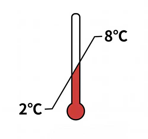
组分(Materials Provided)
| ID | Components | Size |
| RAS175-C01 | Pre-coated Pre-Fusion glycoprotein F0 (RSV) Microplate | 1 plate |
| RAS175-C02 | RSV-F0 Antibody Positive Control | 100 μL |
| RAS175-C03 | RSV-F0 Antibody Negative Control | 100 μL |
| RAS175-C04 | HRP-Conjugated Antibody | 50 μL |
| RAS175-C05 | 10 x Washing Buffer | 50 mL |
| RAS175-C06 | Dilution Buffer | 50 mL |
| RAS175-C07 | Substrate Solution | 12 mL |
| RAS175-C08 | Stop Solution | 7 mL |
产品概述(Product Overview)
Respiratory syncytial virus (RSV) is a highly contagious virus causing severe infection in infants and the elderly. Various approaches are being used to develop an effective RSV vaccine. The RSV fusion (F) subunit, particularly the cleaved trimeric pre-fusion F, is one of the most promising vaccine candidates under development.
To facilitate the RSV-related research, drug trials and vaccine development, a high-throughput assay to measure IgG antibodies against the virus is in urgent need.
应用说明(Application)
The kit is developed for titer measurement of Anti-RSV-F0 Antibody IgG (Pre-Fusion glycoprotein F0) in Human serum.
It is for research use only.
存储(Storage)

原理(Assay Principles)
This assay kit employs a standard indirect-ELISA format, providing a rapid detection of Anti-RSV-F0 antibodies in Human serum by Pre-Fusion glycoprotein F0 (RSV). The Kit consists of Pre-coated Pre-Fusion glycoprotein F0 (RSV) Microplate, Positive Control, Negative Control and HRP-conjugated antibody.
Your experiment will include 4 simple steps:
a) The samples and Control are diluted by Dilution Buffer.Add your sample to the plate.
b) Add the HRP-conjugated antibody diluted by Dliution Buffer to the plate.
c) Wash the plate and add TMB or other colorimetric HRP substrate.
d) Stop the substrate reaction by adding diluted acid. Absorbance (OD) is calculated as the absorbance at 450 nm minus the absorbance at 630 nm to remove background prior to statistical analysis. The OD Value reflects the amount of antibody bound.


 膜杰作
膜杰作 Star Staining


