登录 | 注册    关注公众号  
微信公众号
搜索
 >  Antibody>Prefusion glycoprotein F0/pre-F protein (RSV) >HRV-M720

Monoclonal Anti-HRSV F-hRSV90 Antibody, Human IgG1 (MALS verified)

抗体来源(Source)

Monoclonal Anti-HRSV F-hRSV90 Antibody, Human IgG1 is a chimeric monoclonal antibody recombinantly expressed from HEK293, which combines the variable region of a mouse monoclonal antibody with Human constant domain.

克隆号(Clone)

hRSV90

亚型(Isotype)

Human IgG1 | Human Kappa

偶联(Conjugate)

Unconjugated

抗体类型(Antibody Type)

Recombinant Monoclonal

特异性(Specificity)

This product is a specific antibody specifically reacts with HRSV F.

应用(Application)

ApplicationRecommended Usage
ELISA0.03-313 ng/mL

纯度(Purity)

>90% as determined by SDS-PAGE.

>90% as determined by SEC-MALS.

纯化(Purification)

Protein A purified / Protein G purified

制剂(Formulation)

Supplied as 0.2 μm filtered solution in PBS, pH7.4 with trehalose as protectant.

Contact us for customized product form or formulation.

运输(Shipping)

This product is supplied and shipped with dry ice, please inquire the shipping cost.

存储(Storage)

Please avoid repeated freeze-thaw cycles.

This product is stable after storage at:

  1. The product MUST be stored at -70°C or lower upon receipt;
  2. -70°C for 3 months under sterile conditions.

质量管理控制体系(QMS)

  1. 质量管理体系(ISO, GMP)
  2. 质量优势
  3. 质控流程
 

电泳(SDS-PAGE)

Prefusion glycoprotein F0/pre-F protein (RSV) SDS-PAGE

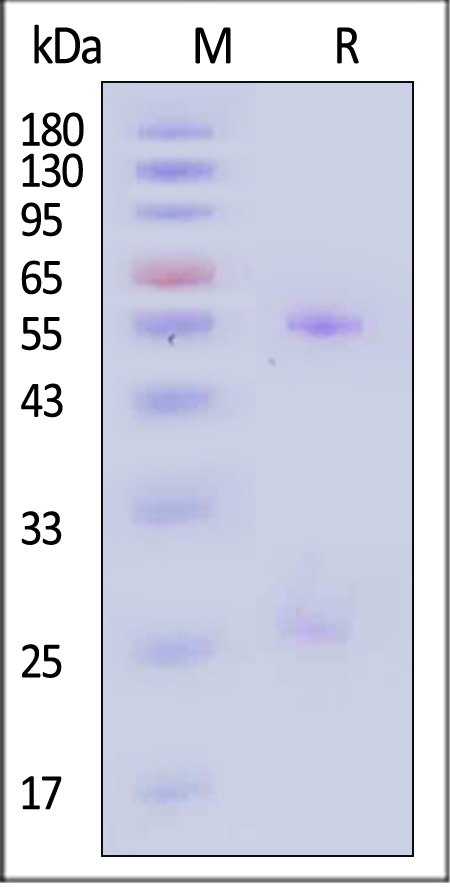
Monoclonal Anti-HRSV F-hRSV90 Antibody, Human IgG1 on SDS-PAGE under reducing (R) condition. The gel was stained with Coomassie Blue. The purity of the protein is greater than 90% (With Star Ribbon Pre-stained Protein Marker).

SEC-MALS

Prefusion glycoprotein F0/pre-F protein (RSV) SEC-MALS

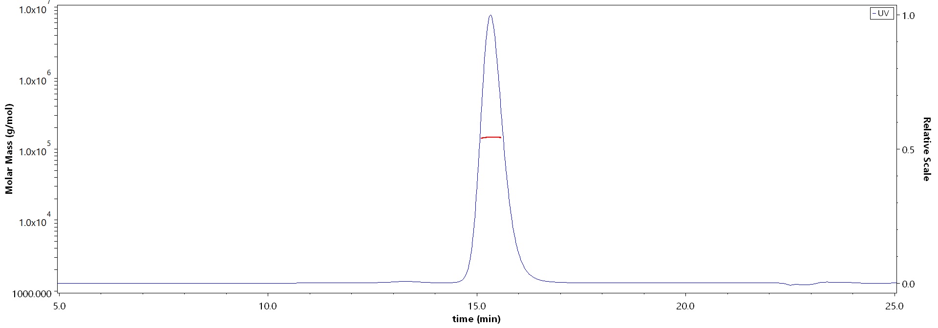
The purity of Monoclonal Anti-HRSV F-hRSV90 Antibody, Human IgG1 (Cat. No. HRV-M720) is more than 90% and the molecular weight of this protein is around 135-165 kDa verified by SEC-MALS.

Report


活性(Bioactivity)-ELISA

Prefusion glycoprotein F0/pre-F protein (RSV) ELISA

Immobilized HRSV (A) Pre-fusion glycoprotein F0, His Tag (Cat. No. RSF-V52H7) at 1 μg/mL (100 μL/well) can bind Monoclonal Anti-HRSV F-hRSV90 Antibody, Human IgG1 (Cat. No. HRV-M720) with a linear range of 0.03-1 ng/mL. HRSV (A) Post-fusion glycoprotein F0, His Tag (Cat. No. RSF-V52H6) is verified not recoginized by Monoclonal Anti-HRSV F-hRSV90 Antibody, Human IgG1 (Cat. No. HRV-M720) in low concentration (QC tested).

Protocol

Prefusion glycoprotein F0/pre-F protein (RSV) ELISA

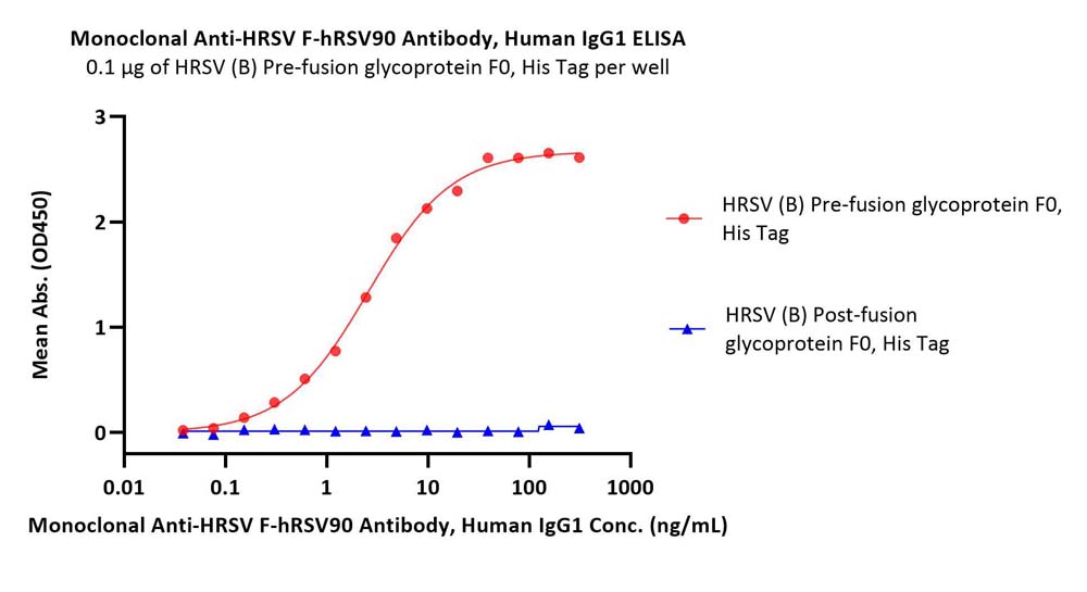
Immobilized HRSV (B) Pre-fusion glycoprotein F0, His Tag (Cat. No. RSF-V52H8) at 1 μg/mL (100 μL/well) can bind Monoclonal Anti-HRSV F-hRSV90 Antibody, Human IgG1 (Cat. No. HRV-M720) with a linear range of 0.03-1 ng/mL. HRSV (B) Post-fusion glycoprotein F0, His Tag (Cat. No. RSF-V52H9) is verified not recoginized by Monoclonal Anti-HRSV F-hRSV90 Antibody, Human IgG1 (Cat. No. HRV-M720) in low concentration (Routinely tested).

Protocol

 
评论(0)
 
ACRO质量管理体系


背景(Background)

Human respiratory syncytial virus (HRSV) is the most common etiological agent of acute lower respiratory tract disease in infants and can cause repeated infections throughout life. The RSV fusion glycoprotein (RSV F) is the principal target of RSV neutralizing antibodies in human sera. The RSV F is a type I viral fusion protein synthesized as inactive, single-chain polypeptides that assemble into trimers. RSV F fuses the viral and host cell membranes by irreversible protein refolding from the labile prefusion conformation to the stable post-fusion conformation. The antibody hRSV90 heavy chain interacts with residues 163–181 and site Ø, while the light chain interacts with residues 163–181 and site II.


前沿进展

Patient outcomes in advanced ovarian cancer treated with an anti-FOLR1 antibody-drug conjugate
Johannet, Flint, Chui et al
Gynecol Oncol (2025) 195, 173-179
Abstract: Mirvetuximab soravtansine-gynx (MIRV) is a FOLR1-binding antibody-drug conjugate (ADC) with a microtubule inhibitor payload. We investigated MIRV's efficacy, toxicity profile, and determinants of resistance in a cohort of patients with recurrent/persistent high FOLR1-expressing high-grade serous ovarian cancer (HGSOC).This retrospective study included 170 patients with recurrent/persistent FOLR1-high (≥75 % of tumor cells with ≥2+ membranous staining intensity) HGSOC who received standard-of-care MIRV monotherapy. We evaluated progression-free survival (PFS) and overall survival (OS) using the Kaplan-Meier method and multivariable Cox proportional hazards models. We classified adverse events using CTCAE v5.0.Overall, median PFS was 3.5 months (95 % CI, 3.0-4.1). However, 22.4 % had PFS ≥6 months and were less likely to have progressed on or within one month of prior taxane-based therapy (P = 0.008). Patients with previous progression on a taxane had worse PFS (HR, 1.69; 95 % CI, 1.19-2.40; P = 0.003) and OS (HR, 2.34; 95 % CI, 1.45-3.77; adjusted P = 0.0005). FOLR1 expression was lower in post-MIRV samples (n = 12; P = 0.005). New or worsening neuropathy was observed in 37.6 % of patients. Among the 34.1 % who experienced ocular toxicity, median onset was 42.5 days. Treatment was discontinued in 5.3 % of patients due to toxicity.MIRV confers meaningful PFS benefit for a subset of individuals with HGSOC. Resistance may be associated with decreased FOLR1 target expression or payload resistance. FOLR1-targeted ADCs with a different payload should be evaluated for patients who progress on MIRV but retain high tumor FOLR1 expression.Copyright © 2025. Published by Elsevier Inc.
Evaluation of Laboratory-Derived Immunohistochemical Assays for Folate Receptor α Expression in Epithelial Ovarian Cancer and Comparison With a Companion Diagnostic
Deutschman, Fulton, Sloss
Arch Pathol Lab Med (2025)
Abstract: The VENTANA FOLR1 (FOLR1-2.1) RxDx (FOLR1 CDx) assay, developed by Roche Tissue Diagnostics, is a Food and Drug Administration-approved immunohistochemical assay intended for use in the assessment of folate receptor α (FRα) expression in formalin-fixed, paraffin-embedded epithelial ovarian, fallopian tube, and primary peritoneal tumor specimens. No published reports have compared the performance of other available FRα antibodies with the approved FOLR1 CDx.To assess the performance of research FRα laboratory-developed tests compared with the FOLR1 CDx.The performance of 6 FRα-targeting antibodies was compared with the approved FOLR1 CDx in normal fallopian tube specimens. Two antibodies were selected for further assessment and compared with the FOLR1 CDx in ovarian tumor specimens.Of the 6 antibodies tested, 4 displayed a lack of specific membrane staining and/or high background, whereas 2 antibodies, produced by Leica Biosystems and Biocare Medical, respectively, exhibited specific and sensitive FRα staining. When assessed for their ability to correctly identify FRα-positive samples (per the FOLR1 CDx label, ≥75% of viable tumor cells with moderate and/or strong membranous staining intensity), both assays overpredicted FRα positivity compared with the FOLR1 CDx in archival ovarian tumor samples.These data highlight the need for caution in antibody selection when developing immunohistochemistry-based assays, as some antibodies failed to cleanly and specifically identify FRα expression. We identified 2 antibodies appropriate for further investigation; however, as developed, these antibodies may overselect patients for treatment with FRα-targeted therapies.© 2025 College of American Pathologists.
Folate receptor 1 is a stemness trait-associated diagnostic and prognostic marker for hepatocellular carcinoma
Shiode, Kodama, Sato et al
Biomark Res (2025) 13 (1), 37
Abstract: Hepatocellular carcinoma (HCC) can be classified into several subtypes based on molecular traits, aiding in prognostic stratification. The subtype with a poor prognosis is often associated with stem/progenitor features. This study focused on identifying circulating biomarkers for aggressive HCC.We searched for secretory proteins whose expression was positively associated with the stem/progenitor markers KRT19, EPCAM, and PROM1 in 2 independent HCC cohorts. Serum folate receptor 1 (FOLR1) levels were measured in 238 chronic liver disease and 247 HCC patients, evaluating their diagnostic and prognostic capabilities.FOLR1 was identified as a secretory protein that was positively correlated with all 3 stem/progenitor markers and a poor prognosis in both the discovery and validation cohorts. Higher FOLR1 expression was detected in tumor than nontumor tissues and was associated with aggressive subtypes, and activation of p53, DNA repair, Myc, E2F, and PI3K/AKT/mTOR pathways. Serum FOLR1 levels correlated with tumoral FOLR1 expression in HCC patients and were significantly elevated compared with those in patients with chronic hepatitis or nonliver disease. Serum FOLR1 levels demonstrated diagnostic performance for HCC comparable to that of alpha-fetoprotein (AFP), and their combination increased the diagnostic accuracy. Elevated serum FOLR1 levels were associated with poor prognosis in HCC patients, regardless of treatment, especially in patients with early-stage disease. The multivariate analysis revealed that the serum FOLR1 level and the Gender, Age, AFP-L3, AFP, and Des-gamma-carboxy prothrombin (GALAD) score were independent predictors of a poor prognosis with their combination further stratifying prognosis.FOLR1 is a stemness-associated biomarker for HCC, with serum levels serving as a diagnostic marker for HCC and a prognostic indicator for early-stage disease.© 2025. The Author(s).
Showing 1-4 of 419 papers.
Powered by BizGenius
 
 
货号/价格
文档
联系电话:
+86 400-682-2521(全国)
010-53681107(北京)
021-50850665(上海)
运输方式
订单邮箱:
order.cn@acrobiosystems.com
技术支持邮箱:
tech.cn@acrobiosystems.com
前沿进展
点击查看详细

消息提示

请输入您的联系方式,再点击提交!

确定